

Intraocular (Uveal) Melanoma
Melanoma of the uveal tract (iris, ciliary body, and choroid), though rare, is the most common primary intraocular malignancy in adults. The mean age-adjusted incidence of uveal melanoma in the United States is approximately 4.3 new cases per million people.
Uveal melanoma is diagnosed mostly at older ages, with a progressively rising, age-specific, incidence rate that peaks near the age of 70 years.
Host susceptibility factors associated with the development of this cancer include:
•Caucasian race. •Light eye color. •Fair skin. •The ability to tan.
In view of these susceptibility factors, numerous observational studies have attempted to explore the relationship between sunlight exposure and risk of uveal melanoma. To date, these studies have found only weak associations or yielded contradictory results. Similarly, there is no consistent evidence that occupational exposure to UV light or other agents is a risk factor for uveal melanoma.
Uveal melanomas differ significantly from cutaneous melanomas. ln one series, 83% of 186 uveal melanomas were found to have a constitutively active somatic mutation in GNAQ or GNA11.
Anatomic Location
Uveal melanomas can arise in the anterior (iris) or the posterior (ciliary body or choroid) uveal tract. Iris melanomas have the best prognosis, whereas melanomas of the ciliary body have the least favorable prognosis. Most uveal tract melanomas originate in the choroid. The ciliary body is less commonly a site of origin, and the iris is the least common. The comparatively low incidence of iris melanomas has been attributed to the characteristic features of these tumors, i.e., they tend to be small, slow growing, and relatively dormant in comparison with their posterior counterparts. Iris melanomas rarely metastasize. Melanomas of the posterior uveal tract generally have a more malignant, histologic appearance; are detected later; and metastasize more frequently than iris melanomas. The typical choroidal melanoma is a brown, elevated, dome-shaped subretinal mass. The degree of pigmentation ranges from dark brown to totally amelanotic.
Most uveal melanomas are initially completely asymptomatic. As the tumor enlarges, it may cause distortion of the pupil (iris melanoma), blurred vision (ciliary body melanoma), or markedly decreased visual acuity caused by secondary retinal detachment (choroidal melanoma). Serous detachment of the retina may occur. If extensive detachment occurs, secondary angle-closure glaucoma occasionally develops. Clinically, several lesions simulate uveal melanoma, including metastatic carcinoma, posterior scleritis, and benign tumors, such as nevi and hemangiomas.
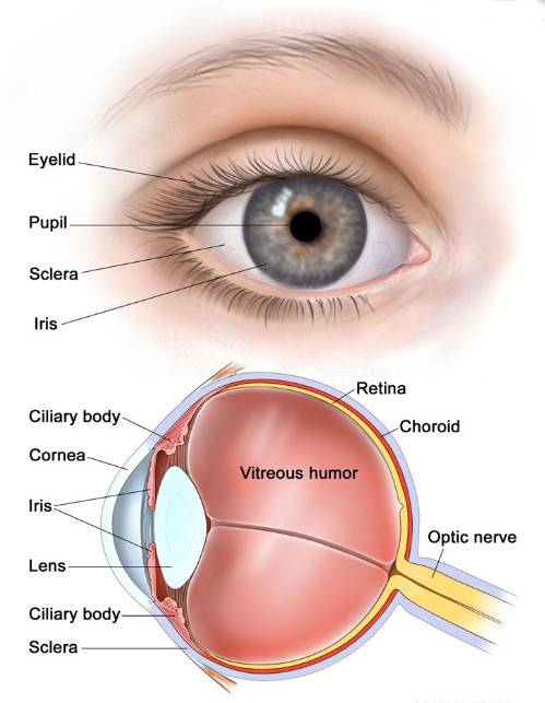
Iris
The iris is the colored area at the front of the eye (the "eye color"). It can be seen through the clear cornea. The pupil is in the center of the iris and it changes size to let more or less light into the eye. Intraocular melanoma of the iris is usually a small tumor that grows slowly and rarely spreads to other parts of the body.
Ciliary body
The ciliary body is a ring of tissue with muscle fibers that change the size of the pupil and the shape of the lens. It is found behind the iris. Changes in the shape of the lens help the eye focus. The ciliary body also makes the clear fluid that fills the space between the cornea and the iris. Intraocular melanoma of the ciliary body is often larger and more likely to spread to other parts of the body than intraocular melanoma of the iris.
Choroid
The choroid is a layer of blood vessels that bring oxygen and nutrients to the eye. Most intraocular melanomas begin in the choroid. Intraocular melanoma of the choroid is often larger and more likely to spread to other parts of the body than intraocular melanoma of the iris.
Diagnosis
Careful examination by an experienced clinician remains the most important test to establish the presence of intraocular melanoma. It is not possible to distinguish a small uveal melanoma from a nevus. Small uveal lesions are often observed for growth to make a diagnosis of melanoma. Clinical findings that may help to identify melanoma include:
•Tumor thickness of more than 2 mm.
•Subretinal fluid.
•Visual symptoms.
•Orange pigment on the tumor surface.
•A tumor margin touching the optic disc.
Ancillary diagnostic testing, including fluorescein angiography and ultrasonography, can be extremely valuable in establishing and/or confirming the diagnosis. In a large, retrospective, single-center series of 2,514 consecutive patients with choroidal nevi, the progression rates to melanoma at 5, 10, and 15 years were 8.6%, 12.8%, and 17.3%, respectively.
Prognostic Factors
A number of factors influence prognosis. The most important factors include the following:
•Cell type. (See Cellular Classification of Intraocular [Uveal] Melanoma section).
•Tumor size.
•Location of the anterior margin of the tumor.
•Degree of ciliary body involvement.
•Extraocular extension.
Several additional microscopic features can affect the prognosis of intraocular melanoma, including:
•Mitotic activity.
•Lymphocytic infiltration.
•Fibrovascular loops (possibly).
Cell type is the most commonly used predictor of outcome following enucleation, with spindle-A cell melanomas carrying the best prognosis and epithelioid cell melanomas carrying the least favorable prognosis. Nevertheless, most tumors have an admixture of cell types, and there is no clear consensus regarding the proportion of epithelioid cells that constitutes designation of a tumor as mixed or epithelioid.
Extraocular extension, recurrence, and metastasis are associated with an extremely poor prognosis, and long-term survival cannot be expected. The 5-year mortality rate associated with metastasis from ciliary body or choroidal melanoma is approximately 30%, compared with a rate of 2% to 3% for iris melanomas.
Cellular Classification of Intraocular (Uveal) Melanoma
Primary intraocular melanomas originate from melanocytes in the uveal tract. Four distinct cellular types are recognized in intraocular melanoma (revised Callender classification):
1.Spindle-A cells (spindle-shaped cells with slender nuclei and lacking visible nucleoli).
2. Spindle-B cells (spindle-shaped cells with larger nuclei and distinct nucleoli).
3. Epithelioid cells (larger polygonal cells with one or more prominent nucleoli).
4. Intermediate cells (similar to but smaller than epithelioid cells).
Most primary intraocular melanomas contain variable proportions of epithelioid, spindle-A, and spindle-B cells (mixed-cell melanomas). Pure epithelioid-cell primary melanomas are infrequent (approximately 3% of cases). In the Collaborative Ocular Melanoma Study, mixed-cell type melanomas predominated (86% of cases).
Staging and Treatment
(See National Cancer Institute (NCI) - Melanoma Treatment PDQ At PubMed)
T1 Tumor limited to the iris.
T2 Tumor confluent with or extending into the ciliary body, choroid, or both.
T3 Tumor confluent with or extending into the ciliary body, choroid, or both, with scleral extension.
T4 Tumor with extrascleral extension.
Iris melanomas have relatively good outcomes with a 5-year survival rate of more than 95%. They are predominantly of the spindle-cell type and are usually smaller in size than posterior melanomas because of earlier detection. Conservative management is generally advocated whenever possible, but surgical intervention may be justified with unequivocal tumor growth or with extensive disease at initial examination.
The management of small choroidal melanomas is controversial, and it is not clear whether treatment of small tumors prevents metastasis. The natural history of small choroidal melanoma is poorly understood. Small, pigmented, choroidal lesions cannot always be differentiated reliably on examination. Growth is a presumed indicator of malignant potential. The likelihood of progression from the time of diagnosis to the time when tumor growth warrants treatment has not been well characterized. Some ophthalmologists advocate observation. This has been justified on several grounds, including the difficulty with establishing a correct diagnosis, the lack of any documented efficacy for globe-conserving treatments, and concerns for severe treatment-related morbidity. Others have advocated earlier therapeutic intervention.
Although patients diagnosed with small choroidal tumors were not eligible for participation in the Collaborative Ocular Melanoma Study (COMS), these patients were offered participation in a prospective follow-up study to evaluate the natural history of small lesions. Two-year and 5-year tumor growth estimates of 21% and 31%, respectively, were reported.[5] Clinical risk factors associated with tumor growth included:
•Increased tumor thickness.
•Presence of subretinal fluid.
•Orange pigmentation.
•Absence of drusen.
•Absence of retinal pigment.
•Margin at the optic disc.
•Epithelial changes surrounding the tumor
No effective method of systemic treatment has been identified for patients with metastatic ocular melanoma. Available clinical trials should be considered as an option for these patients.