Uterine Cervical Cancer
Cervical cancer is the fourth most common cancer in women worldwide, and it has the fourth highest mortality rate among cancers in women.[1] Most cases of cervical cancer are preventable by routine screening and by treatment of precancerous lesions. As a result, most of the cervical cancer cases are diagnosed in women who live in regions with inadequate screening protocols.
Incidence and Mortality
Estimated new cases and deaths from cervical (uterine cervix) cancer in the United States in 2018:
• New cases: 13,240. • Deaths: 4,170.
Anatomy
The uterine cervix is contiguous with the uterine body, and it acts as the opening to the body of the uterus.
The uterine cervix is a cylindrical, fibrous organ that is an average of 3 to 4 cm in length. The portio of the cervix is the part of the cervix that is visible on vaginal inspection.
The opening of the cervix is termed the external os. The os is the beginning of the endocervical canal, which forms the inner aspect of the cervix.
At the upper aspect of the endocervical canal is the internal os, a narrowing of the endocervical canal. The narrowing marks the transition from the cervix to the uterine body. The endocervical canal beyond the internal os is termed the endometrial canal.
The cervix is lined by two types of epithelial cells: squamous cells at the outer aspect, and columnar, glandular cells along the inner canal.
The transition between squamous cells and columnar cells is an area termed the squamo-columnar junction. Most of precancerous and cancerous changes arise in this zone.
Pathogenesis
Cervical carcinoma has its origins at the squamous-columnar junction; it can involve the outer squamous cells, the inner glandular cells, or both.
The precursor lesion is dysplasia: cervical intraepithelial neoplasia (CIN) or adenocarcinoma in situ, which can subsequently become invasive cancer.
This process can be quite slow. Longitudinal studies have shown that in patients with untreated in situ cervical cancer, 30% to 70% will develop invasive carcinoma over a period of 10 to 12 years.
However, in about 10% of patients, lesions can progress from in situ to invasive in a period of less than 1 year.
As it becomes invasive, the tumor breaks through the basement membrane and invades the cervical stroma. Extension of the tumor in the cervix may ultimately manifest as ulceration, exophytic tumor, or extensive infiltration of underlying tissue, including the bladder or rectum.
Prevention of Cervical Cancer
Cervical cancer can often be prevented by having regular screenings to find any precancers and treat them.
Preventing precancers means controlling possible risk factors, such as:
• Delaying first sexual intercourse until the late teens or older
• Limiting the number of sex partners
• Avoiding sexual intercourse with people who have had many partners
• Avoiding sexual intercourse with people who are obviously infected with genital warts or show other symptoms
• Quitting smoking
The HPV vaccine helps prevent cervical cancer caused by HPV.
Gardasil 9 is available in the United States for preventing infection from HPV-16, HPV-18, and 5 other types of HPV linked with cancer.
There were 2 other vaccines previously available in the United States: Cervarix and the original Gardasil. Both of these are no longer available in the United States. However, these vaccines may be in use outside of the United States.
To help prevent cervical cancer, ASCO recommends that girls receive HPV vaccination. Talk with a health care provider about the appropriate schedule for vaccination because it may vary based on many factors, including age and vaccine availability.
Risk Factors
Increasing age is the most important risk factor for most cancers.
Girls younger than 15 years old rarely develop cervical cancer. The risk goes up between the late teens and mid-30s. Women over 40 years of age remain at risk and need to continue having regular cervical cancer screenings, which include both a Pap test and HPV test.
The primary risk factor for cervical cancer is human papillomavirus (HPV) infection.
Research shows that infection with this virus is a risk factor for cervical cancer. Sexual activity with someone who has HPV is the most common way someone gets HPV. There are over 100 different types of HPV, not all of which are linked to cancer. The HPV types that are most frequently associated with cervical cancer are HPV16 and HPV18.
Other risk factors for cervical cancer include the following:
• High parity and HPV infection.
• Smoking cigarettes and HPV infection.
Women who smoke are about twice as likely to develop cervical cancer as women who do not smoke.
• Long-term use of oral contraceptives and HPV infection.
Some research studies suggest that oral contraceptives may be associated with an increase in the risk of cervical cancer. However, more research is needed to understand how oral contraceptive use and the development of cervical cancer are connected.
• Immunosuppression (Immune system deficiency).
Women with lowered immune systems have a higher risk of developing cervical cancer.
• Having first sexual encounter at a young age.
• High number of sexual partners.
• Herpes. Women who have genital herpes have a higher risk of developing cervical cancer.
• Exposure to diethylstilbestrol (DES) in utero.
Women whose mothers were given this drug during pregnancy to prevent miscarriage have an increased risk of developing a rare type of cancer of the cervix or vagina. DES was given for this purpose from about 1940 to 1970. Women exposed to DES should have an annual pelvic examination that includes a cervical Pap test as well as a 4-quadrant Pap test.
• Socioeconomic factors. Cervical cancer is more common among groups of women who are less likely to have access to screening for cervical cancer. Those populations are more likely to include black women, Hispanic women, and American Indian women.
Human papillomavirus (HPV) infection
HPV infection is a necessary step in the development of virtually all precancerous and cancerous lesions. Epidemiologic studies convincingly demonstrate that the major risk factor for development of preinvasive or invasive carcinoma of the cervix is HPV infection, far outweighing other known risk factors.
More than 6 million women in the United States are estimated to be infected with HPV. Transient HPV infection is common, particularly in young women, while cervical cancer is rare. The persistence of an HPV infection leads to increased risk of developing precancerous and cancerous lesions.
The strain of HPV infection is also important in conferring risk. There are multiple subtypes of HPV that infect humans; of these, subtypes 16 and 18 have been most closely associated with high-grade dysplasia and cancer. Studies suggest that acute infection with HPV types 16 and 18 conferred an 11-fold to 16.9-fold risk of rapid development of high-grade CIN. Further studies have shown that infection with either HPV 16 or 18 is more predictive than cytologic screening of high-grade CIN or greater disease, and that the predictive ability is seen for up to 18 years after the initial test.
There are two commercially available vaccines that target anogenital-related strains of HPV. The vaccines are directed towards HPV-naïve girls and young women, and although penetration of the vaccine has been moderate, significant decreases in HPV-related diseases have been documented. (Refer to the PDQ summary on Cervical Cancer Prevention for more information.)
Screening of Cervical Cancer
Screening is used to look for cancer or abnormalities that may become cancerous before you have any symptoms or signs. Scientists have developed, and continue to develop, tests that can be used to screen a person for specific types of cancer before signs or symptoms appear.
The overall goals of cancer screening are to:
• Reduce the number of people who die from the cancer, or completely eliminate deaths from cancer
• Reduce the number of people who develop the cancer
Screening information for cervical cancer
The following tests and procedures may be used to screen for cervical cancer:
• Bimanual pelvic exam. In this examination, the doctor will check a woman’s body for any unusual changes in her cervix, uterus, vagina, ovaries, and other nearby organs. To start, the doctor will look for any changes to the woman’s vulva outside the body and then, using an instrument called a speculum to keep the vaginal walls open, the doctor will look inside the woman’s body. Some of the nearby organs are not visible during this exam, so the doctor will then insert 2 fingers of 1 hand inside the patient’s vagina while the other hand gently presses on the lower abdomen to feel the uterus and ovaries. This exam typically takes a few minutes and is done in an examination room at the doctor’s office.
• HPV test. This test is done on a sample of cells removed from the woman’s cervix, the same sample used for the Pap test. This sample is tested for the strains of HPV most commonly linked to cervical cancer. HPV testing may be done by itself or combined with a Pap test. This test may also be done on a sample of cells collected from a woman’s vagina, which she can collect herself.
• Pap test. The Pap test has been the most common test for early changes in cells that can lead to cervical cancer. This test is also called a Pap smear. A Pap test involves gathering a sample of cells from the cervix. It is often done at the same time as a bimanual pelvic exam. A Pap test may be combined with an HPV test.
• Visual inspection with acetic acid (VIA). VIA is a screening test that can be done with few tools and the naked eye. During VIA, a dilution of white vinegar is applied to the cervix. The health care provider then looks for abnormalities on the cervix, which will turn white when exposed to vinegar. This screening test is very useful in places where access to medical care is limited.
Screening recommendations for cervical cancer
Different organizations have looked at the scientific evidence, risks, and benefits of cervical cancer screening. These groups have developed different screening recommendations for women in the United States who have “average risk” of cervical cancer, meaning these women do not have strong risk factors for this disease.
ASCO recommends that all women receive at least 1 HPV test to screen for cervical cancer in their lifetime. Ideally, women 25 to 65 years old should receive screening with the HPV test every 5 years. Women 65 and older may stop screening if their HPV test results have been mostly negative over the previous 15 years. Sometimes, women who are 65 and older and who have tested positive for HPV may continue screening until they are 70.
Decisions about screening for cervical cancer are becoming increasingly individualized. Sometimes, screening may differ from the recommendations discussed above because of a variety of factors. Such factors include the availability of testing and follow-up options in your area, your personal risk factors, and your health history. It’s important to talk with your health care team or a health care professional knowledgeable in cervical cancer screening about how often you should receive screening and which tests are most appropriate.
Cervical Cancer - Symptoms and Signs
Most women do not have any signs or symptoms of a precancer. In many women with early-stage cervical cancer, symptoms are typically seen. In women with advanced and metastatic cancers, the symptoms may be more severe depending on the tissues and organs to which the disease has spread. The cause of a symptom may be a different medical condition that is not cancer, which is why women need to seek medical care if they have a new symptom that does not go away.
Any of the following could be signs or symptoms of cancer:
• Blood spots or light bleeding between or following periods
• Menstrual bleeding that is longer and heavier than usual
• Bleeding after intercourse, douching, or a pelvic examination
• Increased vaginal discharge
• Pain during sexual intercourse
• Bleeding after menopause
• Unexplained, persistent pelvic and/or back pain
Any of these symptoms should be reported to your doctor. If these symptoms appear, it is important to talk with your doctor about them even if they appear to be symptoms of other, less serious conditions. The earlier precancerous cells or cancer is found and treated, the better the chance that the cancer can be prevented or cured.
Clinical Evaluation
In addition to a physical examination, the following tests may be used to diagnose cervical cancer:
• Pelvic examination. In this examination, the doctor feels a woman’s uterus, vagina, ovaries, cervix, bladder, and rectum to check for any unusual changes. A Pap test is often done at the same time.
• Pap test. During a Pap test, the doctor gently scrapes the outside of the cervix and vagina, taking samples of the cells for testing.
Improved Pap test methods have made it easier for doctors to find cancerous cells. Traditional Pap tests can be hard to read because cells can be dried out, covered with mucus or blood, or clump together on the slide.
• The liquid-based cytology test, often referred to as ThinPrep or SurePath, transfers a thin layer of cells onto a slide after removing blood or mucus from the sample. The sample is preserved, so other tests can be done at the same time, such as the HPV test.
• Computer screening, often called AutoPap or FocalPoint, uses a computer to scan the sample for abnormal cells.
• HPV typing. An HPV test is similar to a Pap test, in which the test is done on a sample of cells from the patient’s cervix. The doctor may test for HPV at the same time as a Pap test or after Pap test results show abnormal changes to the cervix. Certain strains of HPV, such as HPV-16 and HPV-18, are seen more often in women with cervical cancer and may help confirm a diagnosis. If the doctor says the HPV test is “positive,” this means the test found the presence of HPV. Many women have HPV but do not have cervical cancer, so HPV testing alone is not an accurate test for cervical cancer.
Cervical Cancer - Diagnosis
Doctors use many tests to find, or diagnose, cancer. They also do tests to learn if cancer has spread to another part of the body from where it started. If this happens, it is called metastasis. For example, imaging tests can show if the cancer has spread. Imaging tests show pictures of the inside of the body. Doctors may also do tests to learn which treatments could work best.
For most types of cancer, a biopsy is the only sure way for the doctor to know whether an area of the body has cancer. In a biopsy, the doctor takes a small sample of tissue for testing in a laboratory. If a biopsy is not possible, the doctor may suggest other tests that will help make a diagnosis.
If the Pap test showed some abnormal cells and the HPV test is positive, then the doctor may suggest 1 or more of the following diagnostic tests:
• Colposcopy. The doctor may do a colposcopy to check the cervix for abnormal areas. Colposcopy can also be used to help guide a biopsy of the cervix. A special instrument called a colposcope is used. The colposcope magnifies the cells of the cervix and vagina, similar to a microscope. It gives the doctor a lighted, magnified view of the tissues of the vagina and the cervix. The colposcope is not inserted into the woman’s body and the examination is not painful. It can be done in the doctor's office and has no side effects. It can be done on pregnant women.
• Biopsy. A biopsy is the removal of a small amount of tissue for examination under a microscope. Other tests can suggest that cancer is present, but only a biopsy can make a definite diagnosis. A pathologist then analyzes the sample(s). A pathologist is a doctor who specializes in interpreting laboratory tests and evaluating cells, tissues, and organs to diagnose disease. If the lesion is small, the doctor may remove all of it during the biopsy. There are several types of biopsies:
• One common method uses an instrument to pinch off small pieces of cervical tissue.
• Sometimes, the doctor wants to check an area inside the opening of the cervix that cannot be seen during a colposcopy. To do this, the doctor uses a procedure called endocervical curettage (ECC). The doctor uses a small, spoon-shaped instrument called a curette to scrape a small amount of tissue from inside the cervical opening.
• A loop electrosurgical excision procedure (LEEP) uses an electrical current passed through a thin wire hook. The hook removes tissue for examination in the laboratory. A LEEP may also be used to remove a precancer or an early-stage cancer.
• Conization (a cone biopsy) removes a cone-shaped piece of tissue from the cervix. Conization may be done as treatment to remove a precancer or an early-stage cancer.
The first 3 types of biopsy are usually done in the doctor's office using a local anesthetic to numb the area. There may be some bleeding and other discharge. Some women experience discomfort similar to menstrual cramps. Conization is done under a general or local anesthetic and may be done in the doctor's office or the hospital.
Clinical Presentation
Cervical cytology (Pap smear) has been the mainstay of cervical cancer screening since its introduction. However, molecular techniques for the identification of HPV DNA are highly sensitive and specific.
Current screening options include the following:
• Cytology alone.
• Cytology and HPV testing.
HPV testing is suggested when it is likely to successfully triage patients into low- and high-risk groups for a high-grade dysplasia or greater lesion.
HPV DNA tests are unlikely to separate patients with low-grade squamous intraepithelial lesions into those who do and those who do not need further evaluation.
A study of 642 women found that 83% had one or more tumorigenic HPV types when cervical cytologic specimens were assayed by a sensitive (hybrid capture) technique.
The authors of the study and of an accompanying editorial concluded that using HPV DNA testing in this setting does not add sufficient information to justify its cost.
HPV DNA testing has proven useful in triaging patients with atypical squamous cells of undetermined significance to colposcopy and has been integrated into current screening guidelines.
Other studies show that patients with low-risk cytology and high-risk HPV infection with types 16, 18, and 31 are more likely to have CIN or microinvasive histopathology on biopsy.
One method has also shown that integration of HPV types 16 and 18 into the genome, leading to transcription of viral and cellular messages, may predict patients who are at greater risk for high-grade dysplasia and invasive cancer.
For women older than 30 years who are more likely to have persistent HPV infection, HPV typing can successfully triage women into high- and low-risk groups for CIN 3 or worse disease. In this age group, HPV DNA testing is more effective than cytology alone in predicting the risk of developing CIN 3 or worse. Other studies have shown the effectiveness of a primary HPV DNA–screening strategy with cytology triage over the previously used cytology-based screening algorithms.
Abnormal cells in the cervix
Cervical cancer begins when healthy cells on the surface of the cervix change and grow out of control, forming a mass called a tumor. A tumor can be cancerous or benign. A cancerous tumor is malignant, meaning it can spread to other parts of the body. A benign tumor means the tumor will not spread.
At first, the changes in a cell are abnormal, not cancerous. Researchers believe, however, that some of these abnormal changes are the first step in a series of slow changes that can lead to cancer.
Some of the abnormal cells go away without treatment, but others can become cancerous. This phase of the disease is called dysplasia, which is an abnormal growth of cells.
The abnormal cells, sometimes called precancerous tissue, need to be removed to keep cancer from developing. Often, the precancerous tissue can be removed or destroyed without harming healthy tissue, but in some cases, a hysterectomy is needed to prevent cervical cancer.
Treatment of a lesion, which is a precancerous area, depends on the following factors:
• The size of the lesion and the type of changes that have occurred in the cells
• If the woman wants to have children in the future
• The woman's age
• The woman's general health
• The preference of the woman and her doctor
If the precancerous cells change into cancer cells and spread deeper into the cervix or to other tissues and organs, then the disease is called cervical cancer.
Cellular Classification of Cervical Cancer
Squamous cell (epidermoid) carcinoma comprises approximately 90% of cervical cancers, and adenocarcinoma comprises approximately 10% of cervical cancers.
Adenosquamous and small cell carcinomas are relatively rare.
Primary sarcomas of the cervix and primary and secondary malignant lymphomas of the cervix have also been reported.
• Squamous cell carcinoma cancers arise in the cells on the outer surface covering of the cervix.
• Adenocarcinoma cancers arise in the glandular cells that line the lower birth canal.
The squamous and glandular cells meet at the opening of the cervix at the “squamocolumnar junction,” which is the site at which most cervical cancers arise.
Prognostic Factors
The prognosis for patients with cervical cancer is markedly affected by the extent of disease at the time of diagnosis. More than 90% of cervical cancer cases can be detected early through the use of the Pap test and HPV testing.[35] Pap and HPV testing are not performed on approximately 33% of eligible women, which results in a higher-than-expected death rate.
Clinical stage
Clinical stage as a prognostic factor is supplemented by several gross and microscopic pathologic findings in surgically treated patients.
Evidence (clinical stage and other findings):
1. In a large, surgicopathologic staging study of patients with clinical stage IB disease reported by the Gynecologic Oncology Group (GOG) (GOG-49), the factors that most prominently predicted for lymph node metastases and a decrease in disease-free survival were capillary-lymphatic space involvement by tumor, increasing tumor size, and increasing depth of stromal invasion, with the latter being the most important and reproducible.
2. In a study of 1,028 patients treated with radical surgery, survival rates correlated more consistently with tumor volume (as determined by precise volumetry of the tumor) than with clinical or histologic stage.
3. A multivariate analysis of prognostic variables in 626 patients with locally advanced disease (primarily stages II, III, and IV) studied by the GOG identified the following variables that were significant for progression-free interval and survival:
•Periaortic and pelvic lymph node status.
•Tumor size.
•Patient age.
•Performance status.
•Bilateral disease.
•Clinical stage.
The study confirmed the overriding importance of positive periaortic nodes and suggested further evaluation of these nodes in locally advanced cervical cancer. The status of the pelvic nodes was important only if the periaortic nodes were negative. This was also true for tumor size.
In a large series of cervical cancer patients treated by radiation therapy, the incidence of distant metastases (most frequently to the lung, abdominal cavity, liver, and gastrointestinal tract) was shown to increase as the stage of disease increased, from 3% in stage IA to 75% in stage IVA.
A multivariate analysis of factors influencing the incidence of distant metastases showed stage, endometrial extension of tumor, and pelvic tumor control to be significant indicators of distant dissemination.
GOG studies have indicated that prognostic factors vary depending on whether clinical or surgical staging are utilized and with different treatments.
Delay in radiation delivery completion is associated with poorer progression-free survival when clinical staging is used. To-date, stage, tumor grade, race, and age are uncertain prognostic factors in studies utilizing chemoradiation.
Adenocarcinoma vs squamous cell carcinoma
It is controversial whether adenocarcinoma of the cervix carries a significantly worse prognosis than squamous cell carcinoma of the cervix.
Several population-based and retrospective studies show a worse outcome for patients with adenocarcinoma, with an increase in distant metastasis noted, when compared with those with squamous histology.
Reports conflict about the effect of adenosquamous cell type on outcome. One report showed that approximately 25% of apparent squamous tumors have demonstrable mucin production and behave more aggressively than their pure squamous counterparts, suggesting that any adenomatous differentiation may confer a negative prognosis.
Other prognostic factors
• Human immunodeficiency virus (HIV) status: Women with HIV have more aggressive and advanced disease and a poorer prognosis.
• C-myc overexpression: A study of patients with known invasive squamous carcinoma of the cervix found that overexpression of the C-myc oncogene was associated with a poorer prognosis.
• Number of cells in S phase: The number of cells in S phase may also have prognostic significance in early cervical carcinoma.
• HPV-18 DNA: HPV-18 DNA has been found to be an independent adverse molecular prognostic factor. Two studies have shown a worse outcome when HPV-18 was identified in cervical cancers of patients undergoing radical hysterectomy and pelvic lymphadenectomy.
• A polymorphism in the Gamma-glutamyl hydrolase enzyme, which is related to folate metabolism, has been shown to decrease response to cisplatin, and as a result is associated with poorer outcomes.
Follow-up After Treatment
High-quality studies are lacking, and the optimal treatment follow-up for patients after treatment for cervical cancer is unknown. Retrospective studies have shown that patients who recur are most likely to do so within the first 2 years. As a result, most guidelines suggest routine follow-up every 3 to 4 months for the first 2 years, followed by evaluations every 6 months. Most recurrences are diagnosed secondary to new patient symptoms and signs, and the usefulness of routine testing including a Pap smear and chest x-ray is unclear.
Follow-up should be centered around a thorough history and physical examination with a careful review of symptoms; imaging should be reserved for evaluation of a positive finding. Patients should be asked about possible warning signs, including the following:
• Abdominal pain.
• Back pain.
• Painful or swollen leg.
• Problems with urination.
• Cough.
• Fatigue.
The follow-up examination should also screen for possible complications of previous treatment because of the multiple modalities (surgery, chemotherapy, and radiation) that patients often undergo during their treatment.
Staging Procedures
If the biopsy shows that cervical cancer is present, the doctor will refer the woman to a gynecologic oncologist, who may suggest additional tests to see if the cancer has spread beyond the cervix.
• Pelvic examination under anesthesia. In cases where it is necessary for treatment planning, the specialist may re-examine the pelvic area while the patient is under anesthesia to see if the cancer has spread to any organs near the cervix, including the uterus, vagina, bladder, or rectum.
• X-ray. An x-ray is a way to create a picture of the structures inside of the body using a small amount of radiation. An intravenous urography is a type of x-ray that is used to view the kidneys and bladder.
• Computed tomography (CT or CAT) scan. A CT scan creates a 3-dimensional picture of the inside of the body using x-rays taken from different angles. A computer combines these images into a detailed, cross-sectional view that shows any abnormalities or tumors. A CT scan can be used to measure the tumor’s size. Sometimes, a special dye called a contrast medium is given before the scan to provide better detail on the image. This dye can be injected into a patient’s vein or given as a pill to swallow.
• Magnetic resonance imaging (MRI). An MRI uses magnetic fields, not x-rays, to produce detailed images of the body. MRI can be used to measure the tumor’s size. A special dye called a contrast medium is given before the scan to create a clearer picture. This dye can be injected into a patient’s vein or given as a pill to swallow.
• Positron emission tomography (PET) or PET-CT scan. A PET scan is usually combined with a CT scan (see above), called a PET-CT scan. However, you may hear your doctor refer to this procedure just as a PET scan. A PET scan is a way to create pictures of organs and tissues inside the body. A small amount of a radioactive sugar substance is injected into the patient’s body. This sugar substance is taken up by cells that use the most energy. Because cancer tends to use energy actively, it absorbs more of the radioactive substance. A scanner then detects this substance to produce images of the inside of the body.
• Cystoscopy. A cystoscopy is a procedure that allows the doctor to view the inside of the bladder and urethra (the canal that carries urine from the bladder) with a thin, lighted tube called a cystoscope. The person may be sedated as the tube is inserted in the urethra. A cystoscopy is used to determine whether cancer has spread to the bladder.
• Proctoscopy (also called a sigmoidoscopy). A proctoscopy is a procedure that allows the doctor to see the colon and rectum with a thin, lighted, flexible tube called a sigmoidoscope. The person may be sedated as the tube is inserted in the rectum. A proctoscopy is used to see if the cancer has spread to the rectum.
• Laparoscopy. A laparoscopy is a procedure that allows the doctor to see the abdominal area with a thin, lighted, flexible tube called a laparoscope. The person is usually sedated because the tube is inserted through an incision in the body.
Stage Information for Cervical Cancer
Carcinoma of the cervix can spread via local invasion, the regional lymphatics, or bloodstream. Tumor dissemination is generally a function of the extent and invasiveness of the local lesion. While cancer of the cervix generally progresses in an orderly manner, occasionally a small tumor with distant metastasis is seen. For this reason, patients must be carefully evaluated for metastatic disease.
Pretreatment surgical staging is the most accurate method to determine the extent of disease, but there is little evidence to demonstrate overall improved survival with routine surgical staging; the staging is usually performed only as part of a clinical trial. Pretreatment surgical staging in bulky but locally curable disease may be indicated in select cases when a nonsurgical search for metastatic disease is negative. If abnormal nodes are detected by computed tomography (CT) scan or lymphangiography, fine-needle aspiration should be negative before a surgical staging procedure is performed.
Tests and procedures to evaluate the extent of the disease include the following:
• CT scan.
• Positron emission tomography scan.
• Cystoscopy.
• Laparoscopy.
• Chest x-ray.
• Ultrasound.
• Magnetic resonance imaging.
FIGO Stage Groupings and Definitions
The Féderation Internationale de Gynécologie et d’Obstétrique (FIGO) and the American Joint Committee on Cancer have designated staging to define cervical cancer; the FIGO system is most commonly used.
FIGO Staging Systems
I - The carcinoma is strictly confined to the cervix (extension to the uterine corpus should be disregarded).
IA - Invasive cancer identified only microscopically. (All gross lesions even with superficial invasion are Stage IB cancers.) Invasion is limited to measured stromal invasion with a maximum depth of 5 mmb and no wider than 7 mm.
IA1: Measured invasion of stroma ≤3.0 mm in depth and ≤7.0 mm width.
IA2: Measured invasion of stroma >3.0 mm and < 5.0 mm in depth and ≤ 7 mm width.
IB - Clinical lesions confined to the cervix or preclinical lesions greater than stage IA.
IB1: Clinical lesions no greater than 4 cm in size.
IB2: Clinical lesions >4 cm in size.
II - The carcinoma extends beyond the uterus but not extended onto the pelvic wall or to the lower third of the vagina.
IIA - Involvement of up to the upper 2/3 of the vagina. No obvious parametrial involvement.
IIA1: Clinically visible lesion ≤4.0 cm.
IIA2: Clinically visible lesion >4.0 cm.
IIB - Obvious parametrial involvement but not onto the pelvic sidewall.
III - The carcinoma has extended onto the pelvic sidewall. On rectal examination, there is no cancer-free space between the tumor and pelvic sidewall. The tumor involves the lower third of the vagina. All cases of hydronephrosis or nonfunctioning kidney should be included unless they are known to be due to other causes.
IIIA - Involvement of the lower vagina but no extension onto pelvic sidewall.
IIIB - Extension onto the pelvic sidewall, or hydronephrosis/non-functioning kidney
IV - The carcinoma has extended beyond the true pelvis or has clinically involved the mucosa of the bladder and/or rectum.
IVA - Spread to adjacent pelvic organs.
IVB - Spread to distant organs.
Treatment Option Overview
Patterns-of-care studies clearly demonstrate the negative prognostic effect of increasing tumor volume and spread pattern.
Treatment, therefore, may vary within each stage as the individual stages are currently defined by Féderation Internationale de Gynécologie et d’Obstétrique (FIGO).
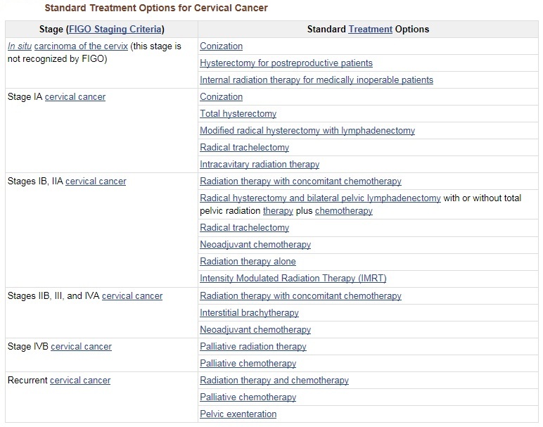
Chemoradiation Therapy
Five randomized, phase III trials (GOG-85, RTOG-9001, GOG-120, GOG-123, and SWOG-8797) have shown an overall survival advantage for cisplatin-based therapy given concurrently with radiation therapy, while one trial examining this regimen demonstrated no benefit. The patient populations in these studies included women with FIGO stages IB2 to IVA cervical cancer treated with primary radiation therapy and women with FIGO stages I to IIA disease who were found to have poor prognostic factors (metastatic disease in pelvic lymph nodes, parametrial disease, or positive surgical margins) at the time of primary surgery.
• Although the positive trials vary in terms of the stage of disease, dose of radiation, and schedule of cisplatin and radiation, the trials demonstrate significant survival benefit for this combined approach. The risk of death from cervical cancer was decreased by 30% to 50% with the use of concurrent chemoradiation therapy.
• Based on these results, strong consideration should be given to the incorporation of concurrent cisplatin-based chemotherapy with radiation therapy in women who require radiation therapy for treatment of cervical cancer.
• Other studies have validated these results.
Surgery and Radiation Therapy
Surgery and radiation therapy are equally effective for early stage, small-volume disease. Younger patients may benefit from surgery to preserve the ovaries and avoid vaginal atrophy and stenosis.
Therapy for patients with cancer of the cervical stump is effective and yields results that are comparable with those seen in patients with an intact uterus.
In Situ Cervical Cancer Treatment
Consensus guidelines have been issued for managing women with cervical intraepithelial neoplasia or adenocarcinoma in situ.
Properly treated, tumor control of in situ cervical carcinoma should be nearly 100%.
Either expert colposcopic-directed biopsy or cone biopsy is required to exclude invasive disease before therapy is undertaken.
A correlation between cytology and colposcopic-directed biopsy is also necessary before local ablative therapy is done.
Unrecognized invasive disease treated with inadequate ablative therapy may be the most common cause of failure.
Failure to identify the disease, lack of correlation between the Pap smear and colposcopic findings, adenocarcinoma in situ, or extension of disease into the endocervical canal makes a laser, loop, or cold-knife conization mandatory.
The choice of treatment depends on the extent of disease and several patient factors, including age, cell type, desire to preserve fertility, and medical condition.
Standard Treatment Options for In Situ Cervical Cancer
1. Conization.
◦ Cold-knife conization (scalpel).
◦ Loop electrosurgical excision procedure (LEEP).
◦ Laser therapy.
2. Hysterectomy for postreproductive patients.
3. Internal radiation therapy for medically inoperable patients.
Hysterectomy is the standard treatment for patients with adenocarcinoma in situ. The disease, which originates in the endocervical canal, may be more difficult to completely excise with a conization procedure. Conization may be offered to select patients with adenocarcinoma in situ who desire future fertility.
Conization
When the endocervical canal is involved, laser or cold-knife conization may be used for selected patients to preserve the uterus, avoid radiation therapy, and more extensive surgery.
In selected cases, the outpatient LEEP may be an acceptable alternative to cold-knife conization. This procedure requires only local anesthesia and obviates the risks associated with general anesthesia for cold-knife conization. However, controversy exists about the adequacy of LEEP as a replacement for conization; LEEP is unlikely to be sufficient for patients with adenocarcinoma in situ.
Evidence (conization using LEEP):
1. A trial comparing LEEP with cold-knife cone biopsy showed no difference in the likelihood of complete excision of dysplasia.
2. Two case reports suggested that the use of LEEP in patients with occult invasive cancer led to an inability to accurately determine depth of invasion when a focus of the cancer was transected.
Hysterectomy for postreproductive patients
Hysterectomy is standard therapy for women with cervical adenocarcinoma in situ, because of the location of the disease in the endocervical canal and the possibility for skip lesions in this region, making margin status a less reliable prognostic factor.
However, the effect of hysterectomy compared with conservative surgical measures on mortality has not been studied.
Hysterectomy may be performed for squamous cell carcinoma in situ if conization is not possible because of previous surgery, or if positive margins are noted after conization therapy.
Hysterectomy is not an acceptable front-line therapy for squamous carcinoma in situ.
Internal radiation therapy for medically inoperable patients
For medically inoperable patients, a single intracavitary insertion with tandem and ovoids for 5,000 mg hours (80 Gy vaginal surface dose) may be used.
Stage IA Cervical Cancer Treatment
Standard Treatment Options for Stage IA1 Cervical Cancer
1. Conization.
2. Total hysterectomy.
Conization
If the depth of invasion is less than 3 mm, no vascular or lymphatic channel invasion is noted, and the margins of the cone are negative, conization alone may be appropriate in patients who wish to preserve fertility.
Total hysterectomy
If the depth of invasion is less than 3 mm, which is proven by cone biopsy with clear margins, no vascular or lymphatic channel invasion is noted, and the frequency of lymph-node involvement is sufficiently low, lymph-node dissection at the time of hysterectomy is not required.
Oophorectomy is optional and should be deferred for younger women.
Standard Treatment Options for Stage IA2 Cervical Cancer
Modified radical hysterectomy with lymphadenectomy
For patients with tumor invasion between 3 mm and 5 mm, modified radical hysterectomy with pelvic-node dissection has been recommended because of a reported risk of lymph-node metastasis of as much as 10%.
Radical hysterectomy with node dissection may also be considered for patients for whom the depth of tumor invasion was uncertain because of invasive tumor at the cone margins.
Other Treatment Options
1. Radical trachelectomy.
2. Intracavitary radiation therapy.
Radical trachelectomy
Patients with stages IA2 to IB disease who desire future fertility may be candidates for radical trachelectomy.
In this procedure, the cervix and lateral parametrial tissues are removed, and the uterine body and ovaries are maintained.
Most centers utilize the following criteria for patient selection:
• Desire for future pregnancy.
• Age younger than 40 years.
• Presumed stage IA2 to IB1 disease and a lesion size no greater than 2 cm.
• Preoperative magnetic resonance imaging that shows a margin from the most distal edge of the tumor to the lower uterine segment.
• Squamous, adenosquamous, or adenocarcinoma cell types.
Intraoperatively, the patient is assessed in a manner similar to a radical hysterectomy; the procedure is aborted if more advanced disease than expected is encountered.
The margins of the specimen are also assessed at the time of surgery, and a radical hysterectomy is performed if inadequate margins are obtained.
radiation therapy
Intracavitary radiation therapy is a treatment option when palliative treatment is appropriate because of other medical conditions and for women who are not surgical candidates.
If the depth of invasion is less than 3 mm and no capillary lymphatic space invasion is noted, and the frequency of lymph-node involvement is sufficiently low, external-beam radiation therapy is not required.
One or two insertions with tandem and ovoids for 6,500 mg to 8,000 mg hours (100–125 Gy vaginal surface dose) are recommended.