Hodgkin Lymphoma
Hodgkin lymphoma, which used to be called Hodgkin’s disease, is one of many types of cancer that develops in the lymphatic system.
Hodgkin lymphoma most commonly affects lymph nodes in the neck or in the area between the lungs and behind the breastbone. It can also begin in groups of lymph nodes under an arm, in the groin, or in the abdomen or pelvis.
If Hodgkin lymphoma spreads, it may spread to the spleen, liver, bone marrow, or bone. Hodgkin lymphoma can spread to other parts of the body, but this is unusual. (ASCO)
Incidence and Mortality
Estimated new cases and deaths from HL in the United States in 2018:
• New cases: 8,500. • Deaths: 1,050.
More than 75% of all newly diagnosed patients with adult HL can be cured with combination chemotherapy and/or radiation therapy.
National mortality has fallen more rapidly for adult HL than for any other malignancy over the last 5 decades
Prognosis and Survival Factors
Prognosis for a given patient depends on several factors. The most important factors are the presence or absence of systemic symptoms, the stage of disease, presence of large masses, and the quality and suitability of the treatment administered. Other important factors are age, sex, erythrocyte sedimentation rate, extent of abdominal involvement, hematocrit, and absolute number of nodal sites of involvement.
HL is the main cause of death over the first 15 years after treatment. By 15 to 20 years after therapy, the cumulative mortality from a second malignancy will exceed the cumulative mortality from HL.
Risk Factors
The exact cause of Hodgkin lymphoma is not known, but the following factors may raise a person’s risk of developing Hodgkin lymphoma:
• Age. People between the ages of 15 and 40 and people older than 55 are more likely to develop Hodgkin lymphoma.
• Gender. In general, men are slightly more likely to develop Hodgkin lymphoma than women, although the nodular sclerosis subtype is more common in women.
• Family history. Brothers and sisters of people with Hodgkin lymphoma have a higher chance of developing the disease, although the increase in risk is small.
• Virus exposure. The Epstein-Barr virus (EBV) causes infectious mononucleosis, often called "mono." Nearly all adult Americans and many others around the world have an EBV infection. About 20% to 25% of people with cHL in the United States have lymphoma cells that test positive for EBV. Although a person’s immune system response to an infection with EBV may be important in the development of Hodgkin lymphoma, doctors still don’t understand why, when so many people have been infected with EBV, relatively very few people ever develop Hodgkin lymphoma. People with HIV also have a higher risk of developing Hodgkin lymphoma, particularly lymphocyte-depleted Hodgkin lymphoma.
Cellular Classification of Adult HL
Pathologists currently use the World Health Organization (WHO) modification of the Revised European-American Lymphoma (REAL) classification for the histologic classification for adult Hodgkin lymphoma (HL).
WHO/REAL classification
• Classical HL.
◦ Nodular sclerosis HL.
◦ Mixed-cellularity HL.
◦ Lymphocyte depletion HL.
◦ Lymphocyte-rich classical HL.
• Nodular lymphocyte–predominant HL.
Among 10,019 patients who underwent central expert pathology review for the German Hodgkin Study Group, 84 patients (<1%) were identified as having lymphocyte-depleted classical HL. These patients present with more advanced-stage HL and usually with B symptoms.
Nodular Lymphocyte–Predominant HL
Nodular lymphocyte–predominant HL (NLPHL) is a clinicopathologic entity of B-cell origin that is distinct from classic HL. The typical immunophenotype for lymphocyte-predominant disease is CD15-, CD20+, CD30-, CD45+, while the profile for classic HL is CD15+, CD20-, CD30+, CD45-.
Patients with lymphocyte-predominant disease have earlier-stage disease, longer survival, and fewer treatment failures than those with classic HL. Despite a usually favorable prognosis, there is a tendency for histologic transformation to diffuse large B-cell lymphoma or T-cell–rich large B-cell lymphoma in approximately 10% of patients by 10 years.
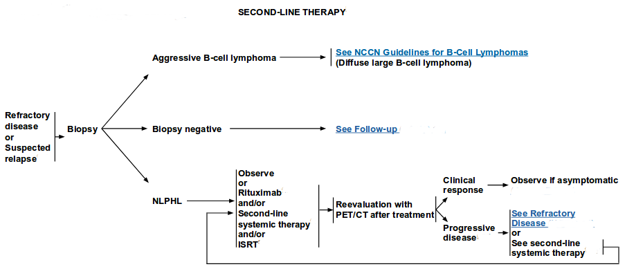
This propensity of NLPHL to transform to aggressive B-cell lymphoma underscores the importance of long-term follow-up and re-biopsy at relapse.
Lymphocyte-predominant HL is usually diagnosed in asymptomatic young males with cervical or inguinal lymph nodes but usually without mediastinal involvement. Based on retrospective analyses spanning several decades and because of the rarity of this histology, limited-field radiation therapy is the most common treatment approach for patients with early-stage disease.
The REAL Classification of Lymphoid Neoplasms proposed separating Nodular lymphocyte–predominant HL (NLPHL) (CD15-, CD20+, CD30-) from lymphocyte-rich classical HL (CD15+, CD20-, CD30+), on the basis of these immunophenotypic differences. The largest retrospective report of 426 cases showed no significant difference in clinical response or outcome to standard therapies for these two subgroups.[Level of evidence: 3iiiA]
Of interest, with a median follow-up of 7 to 8 years, more patients died of treatment-related toxic effects (acute and long-term) than from Hodgkin recurrence. Limitation of radiation dose and fields and avoidance of leukemogenic chemotherapeutic agents, along with watchful waiting policies, should be investigated for these subgroups. For patients with advanced-stage NLPHL, chemotherapy regimens designed for patients with non-HLs may be preferred, based on two retrospective reviews and a phase II study.][Level of evidence: 3iiiDii]
Rituximab had a 100% response rate in a phase II trial of 39 previously untreated and relapsed NLPHL patients. With a median follow-up of 9.8 years, the median PFS was 3.0 years for patients who received rituximab induction only and 5.6 years for patients who received rituximab induction plus rituximab maintenance.[Level of evidence: 3iiiDiii] With induction only, 9 of 23 patients relapsed with an aggressive B-cell lymphoma
Types of Hodgkin lymphoma
The American Joint Committee on Cancer (AJCC) recognizes 2 major categories of Hodgkin lymphoma: classical Hodgkin lymphoma, which is divided into 4 subtypes based on the appearance of the lymph node structure and cells, and nodular lymphocyte-predominant Hodgkin lymphoma.
Classical Hodgkin lymphoma (cHL)
cHL is the most common type of Hodgkin lymphoma. About 95% of cases of Hodgkin lymphoma fall into the cHL category. cHL is diagnosed when characteristic abnormal lymphocytes, known as Reed-Sternberg cells, are found. cHL is divided into 4 subtypes:
• Nodular sclerosis Hodgkin lymphoma. Nodular sclerosis Hodgkin lymphoma is the most common type of cHL. It affects up to 80% of people diagnosed with cHL. Nodular sclerosis Hodgkin lymphoma is most common in young adults, especially women. In addition to Reed-Sternberg cells, there are bands of connective tissue (called fibrosis) found in the lymph node. The presence of these bands can help diagnose this type of Hodgkin lymphoma. This type of lymphoma often affects the lymph nodes in the central part of the chest, called the mediastinum.
• Lymphocyte-rich classical Hodgkin lymphoma. About 6% of people with cHL are diagnosed with lymphocyte-rich classical Hodgkin lymphoma. It is more common in men and usually affects areas other than the mediastinum. In addition to Reed-Sternberg cells, the lymph node tissue contains many normal lymphocytes.
• Mixed cellularity Hodgkin lymphoma. This subtype of cHL occurs most often in older adults. It commonly develops in the abdomen and carries many different cell types, including large numbers of Reed-Sternberg cells.
• Lymphocyte-depleted Hodgkin lymphoma. Lymphocyte-depleted Hodgkin lymphoma is the least common subtype of cHL. Only about 1% of people with cHL have this subtype. It is most common in older adults; people with the human immunodeficiency virus (HIV), the virus that causes autoimmune deficiency syndrome (AIDS); and people in non-industrialized countries. The lymph node contains almost all Reed-Sternberg cells.
Nodular lymphocyte-predominant Hodgkin lymphoma
About 5% of people with Hodgkin lymphoma have nodular lymphocyte-predominant Hodgkin lymphoma. It often develops in the lymph nodes in the neck, groin, or armpit. It is most common in younger patients.
Nodular lymphocyte-predominant Hodgkin lymphoma is more similar to B-cell non-Hodgkin lymphoma. People with this type of Hodgkin lymphoma have large cells in the affected area called “popcorn cells” or “LP cells” that have a marker called CD20 on their surface. CD20 is a protein that is usually found in people diagnosed with B-cell non-Hodgkin lymphoma.
Nodular lymphocyte-predominant Hodgkin lymphoma is often treated differently from cHL. Some people with nodular lymphocyte-predominant Hodgkin lymphoma do not need treatment right away, while others may benefit from a treatment plan that includes radiation therapy, chemotherapy, or a monoclonal antibody called rituximab (Rituxan).
People with nodular lymphocyte-predominant Hodgkin lymphoma tend to have a very good prognosis. This means the treatment, if needed, has a very good chance of being successful and helps the patient recover. However, a small number of patients with nodular lymphocyte-predominant Hodgkin lymphoma may develop a more aggressive type of non-Hodgkin lymphoma called diffuse large B-cell lymphoma through a process called transformation.
Stage Information for Adult Hodgkin Lymphoma
Clinical staging for patients with Hodgkin lymphoma (HL) includes a history, physical examination, laboratory studies (including sedimentation rate), and thoracic and abdominal/pelvic computerized tomographic (CT) scans with or without positron emission tomography (PET).
PET scans combined with CT scans have become the standard imaging for clinical staging.
A prospective, multinational study of 260 newly diagnosed patients with advanced-stage HL obtained PET scans at baseline and after two cycles (four doses) of doxorubicin plus bleomycin plus vinblastine plus dacarbazine (ABVD); with a median follow-up of 2.2 years, the 2-year progression-free survival (PFS) was 12.8% with a positive PET scan after two cycles and 95% with a negative PET scan after two cycles (P < .0001).
In a prospective trial of BEACOPP-based therapy—includes the drugs bleomycin, etoposide, doxorubicin hydrochloride, cyclophosphamide, vincristine sulfate, procarbazine, and prednisone—for previously untreated patients with advanced-stage HL, patients with residual abnormalities measuring 2.5 cm or more received a PET scan at the end of therapy.
A negative PET scan predicted no progression or relapse within 1 year for 94% of patients (confidence interval, 91%–97%).
Whether consolidation with radiation therapy can be omitted for PET-negative patients must await overall survival data at 5 years.
Only further prospective studies that compare a PET response–adapted strategy versus standard therapy without alteration can assess whether improved outcomes can be achieved by altering the therapeutic strategy based on PET scan results.
In two prospective randomized trials encompassing 1,739 patients with clinical stage I or II HL, acute relapse rates were lower with combined modality therapy, even for patients with a negative interim PET scan.
These trials do not support using interim PET scans to determine the value of radiation therapy for early-stage disease.
Both studies showed that patients with early-stage disease and a negative PET scan after two or three cycles of ABVD had a very good prognosis (PFS exceeding 90% at 3 years in one of the studies) with or without consolidation radiation therapy.
Neither of these studies has been followed long enough (>10 years) to assess long-term toxicities or treatment-related mortality.
Bone marrow involvement occurs in 5% of patients; biopsy may be indicated in the presence of constitutional B symptoms or anemia, leukopenia, or thrombocytopenia.
In a retrospective review and meta-analysis of 955 patients in nine studies, fewer than 2% of patients with a positive bone marrow biopsy had only stage I or II disease on PET-CT scans; omission of the bone marrow biopsy for PET-CT–designated early-stage patients did not change treatment selection.
Staging laparotomy is no longer recommended and should be considered only when the results will allow substantial reduction in treatment. Staging laparotomy should not be done in patients who require chemotherapy. If the laparotomy is required for treatment decisions, the risks of potential morbidity should be considered. The staging classification that is currently used for HL was adopted in 1971 at the Ann Arbor Conference with some modifications 18 years later from the Cotswolds meeting.
Subclassification of stage
Stages I, II, III, and IV adult HL can be subclassified into A and B categories: B for those with defined general symptoms and A for those without B symptoms. The B designation is given to patients with any of the following symptoms:
• Unexplained loss of more than 10% of body weight in the 6 months before diagnosis.
• Unexplained fever with temperatures above 38°C.
• Drenching night sweats.
[Note: The most significant B symptoms are fevers and weight loss. Night sweats alone do not confer an adverse prognosis. Pruritus as a systemic symptom remains controversial and is not considered a B symptom in the Ann Arbor staging system. This symptom is hard to define quantitatively and uniformly, but when it is recurrent, generalized, and otherwise unexplained, and when it ebbs and flows parallel to disease activity, it may be the equivalent of a B symptom.]
The designation E is used when well-localized extranodal lymphoid malignancies arise in or extend to tissues beyond, but near, the major lymphatic aggregates. Stage IV refers to disease that is diffusely spread throughout an extranodal site, such as the liver. If pathologic proof of involvement of one or more extralymphatic sites has been documented, the symbol for the site of involvement, followed by a plus sign (+), is listed.
Table 1. Notations for Identifying Sites
N = nodes
H = liver
L = lung
M = bone marrow
S = spleen
P = pleura
O = bone
D = skin
Current practice is to assign a clinical stage (CS) based on the findings of the clinical evaluation and a pathologic stage (PS) based on the findings of invasive procedures.
For example, a patient who has disease in the chest and neck, systemic symptoms, and a negative lymphangiogram might be found at laparotomy to have involvement of the spleen, liver, and bone marrow. Thus, the precise stage of such a patient would be CS IIB, PS IVB (S+)(H+)(M+).
The AJCC has designated staging using the Ann Arbor classification system to define adult Hodgkin lymphoma.
| Stage | Prognostic Groups |
| I | Involvement of a single lymphatic site (i.e., nodal region, Waldeyer ring, thymus or spleen) (I). |
| Localized involvement of a single extralymphatic organ or site in the absence of any lymph node involvement (IE) (rare in Hodgkin lymphoma). | |
| II | Involvement of ≥2 lymph node regions on the same side of the diaphragm (II). |
| Localized involvement of a single extralymphatic organ or site in association with regional lymph node involvement with or without involvement of other lymph node regions on the same side of the diaphragm (IIE). | |
| The number of regions involved may be indicated by an arabic numeral, as in, for example, II3. | |
| III | Involvement of lymph node regions on both sides of the diaphragm (III), which also may be accompanied by extralymphatic extension in association with adjacent lymph node involvement (IIIE) or by involvement of the spleen (IIIS) or both (IIIE, S). |
| Splenic involvement is designated by the letter S. | |
| IV | Diffuse or disseminated involvement of one or more extralymphatic organs, with or without associated lymph node involvement. |
| Isolated extralymphatic organ involvement in the absence of adjacent regional lymph node involvement, but in conjunction with disease in distant site(s). | |
| Stage IV includes any involvement of the liver or bone marrow, lungs (other than by direct extension from another site), or cerebrospinal fluid. |
Massive mediastinal disease has been defined by the Cotswolds meeting as a thoracic ratio of maximum transverse mass diameter of 33% or more of the internal transverse thoracic diameter measured at the T5/6 intervertebral disc level on chest radiography.
Some investigators have designated a lymph node mass measuring 10 cm or more in greatest dimension as massive disease.
Other investigators use a measurement of the maximum width of the mediastinal mass divided by the maximum intrathoracic diameter.
Many investigators and many new clinical trials employ a clinical staging system that divides patients into three major groups that are also useful for the practicing physician:
• Early favorable: Clinical stage I or II without any risk factors.
• Early unfavorable: Clinical stage I or II with one or more of the following risk factors:
◦ Large mediastinal mass (>33% of the thoracic width on the chest x-ray, ≥10 cm on CT scan).
◦ Extranodal involvement.
◦ Elevated erythrocyte sedimentation rate (>30 mm/h for B stage, >50 mm/h for A stage).
◦ Three or more lymph node areas' involvement.
◦ B symptoms.
• Advanced: For patients with advanced-stage HL, the International Prognostic Factors Project has developed an International Prognostic Index with a prognostic score that is based on the following seven adverse factors:
◦ Albumin level of <4.0 g/dL.
◦ Hemoglobin level of <10.5 g/dL.
◦ Male sex.
◦ Age of ≥45 years.
◦ Stage IV disease.
◦ White blood cell (WBC) count of ≥15,000/mm3.
◦ Absolute lymphocytic count of <600/mm3 or a lymphocyte count that was <8% of the total WBC count.
Other prognostic factors that are considered, especially for early-stage Hodgkin lymphoma, include:
• A higher ESR (see Diagnosis) is associated with a poorer prognosis.
• People with lymphocyte-predominant Hodgkin lymphoma, nodular sclerosis Hodgkin lymphoma, and lymphocyte-rich classical Hodgkin lymphoma have a better prognosis, compared with other subtypes of Hodgkin lymphoma.
• A mediastinal lymph node mass, located in the center of the chest, that is larger than 10 centimeters (cm) is associated with a poorer prognosis. Small mediastinal masses are not associated with a poorer prognosis.
• Having a high number of involved lymph node sites is associated with a poorer prognosis
(ASCO)
Treatment Option Overview for Adult Hodgkin Lymphoma
Drug combinations described in this section include the following:
• ABVD: doxorubicin, bleomycin, vinblastine, and dacarbazine.
• BEACOPP: bleomycin, etoposide, doxorubicin, cyclophosphamide, vincristine, procarbazine, and prednisone.
• MOPP: mechlorethamine, vincristine, procarbazine, and prednisone.
After initial clinical staging for Hodgkin lymphoma (HL), patients with obvious stage III or IV disease, bulky disease (defined as a 10 cm mass or mediastinal disease with a transverse diameter exceeding 33% of the transthoracic diameter), or the presence of B symptoms will require combination chemotherapy with or without additional radiation therapy.
Patients with nonbulky stage IA or IIA disease are considered to have clinical early-stage disease. These patients are candidates for chemotherapy, combined modality therapy, or radiation therapy alone. Staging laparotomy is no longer recommended because it may not alter management and does not enhance ultimate outcome. When chemotherapy alone or combined modality therapy is applied, laparotomy is not required.
Radiation Therapy
In adult HL, the appropriate dose of radiation alone is 25 Gy to 30 Gy to clinically uninvolved sites and 35 Gy to 44 Gy to regions of initial nodal involvement.
These recommendations are often modified in pediatric or advanced-staged adult patients who also receive chemotherapy.
Treatment is usually delivered to the neck, chest, and axilla (mantle field) and then to an abdominal field to treat para-aortic nodes and the spleen (splenic pedicle). In some patients, pelvic nodes are treated with a third field.
The three fields constitute total nodal radiation therapy. In some cases, the pelvic and para-aortic nodes are treated in a single field called an inverted Y. In patients with a favorable prognosis, treatment of the pelvic lymph nodes is frequently omitted, since fertility can be preserved without affecting relapse-free survival.
Second Malignancies
Acute nonlymphocytic leukemia may occur in patients treated with combined modality therapy or with combination chemotherapy alone, especially with increasing exposure to alkylating agents.
At 10 years after therapy with regimens containing MOPP, the risk of acute myelogenous leukemia (AML) is approximately 3%, with the peak incidence occurring 5 to 9 years after therapy.
The risk of acute leukemia at 10 years following therapy with ABVD appears to be less than 1%.
A population-based study of more than 35,000 survivors during a 30-year time span identified 217 patients who developed AML; the excess absolute risk is significantly higher (9.9 vs. 4.2 after 1984, P < .001) for older patients (i.e., >35 years at diagnosis) versus younger survivors.
An increase in second solid tumors has also been observed, especially cancers of the lung, breast, thyroid, bone/soft tissue, stomach, esophagus, colon and rectum, uterine cervix, head and neck, and mesothelioma.
These tumors occur primarily after radiation therapy or with combined modality treatment, and approximately 75% occur within radiation ports.
At a 15-year follow-up, the risk of second solid tumors (cumulative incidence of a second cancer) is approximately 13%; at a 20-year follow-up, the risk is approximately 17%; at a 25-year follow-up, the risk is approximately 22%; and at a 40-year follow-up, the risk is approximately 48%.
In a cohort of 18,862 5-year survivors from 13 population-based registries, the younger patients had elevated risks for breast, colon, and rectal cancer for 10 to 25 years before the age when routine screening would be recommended in the general population. Even with involved-field doses of 15 Gy to 25 Gy, sarcomas, breast cancers, and thyroid cancers occurred with similar incidence in young patients receiving higher-dose radiation.
Lung cancer is seen with increased frequency, even after chemotherapy alone, and the risk of this cancer is increased with cigarette smoking.
In a retrospective Surveillance, Epidemiology, and End Results (SEER) analysis, stage-specific survival was decreased by 30% to 60% in HL survivors compared with patients with de novo non-small cell lung cancer.
Breast cancer is seen with increased frequency after radiation therapy or combined modality therapy. The risk appears greatest for women treated with radiation before age 30 years and especially close to menarche, and the incidence increases substantially after 15 years of follow-up.
In two case control studies of 479 patients who developed breast cancer after therapy for HL, cumulative absolute risks for developing breast cancer were calculated as a function of radiation therapy dose and the use of chemotherapy.
With a 30-year to 40-year follow-up, cumulative absolute risks of breast cancer with exposure to radiation range from 8.5% to 39.6%, depending on the age at diagnosis.
A family history of breast cancer or ovarian cancer does not confer a greater increased risk than that of radiation therapy for one of these cohorts. These cohort studies show a continued increase in cumulative excess risk of breast cancer beyond 20 years of follow-up.
In a nested case control study and subsequent cohort study, patients who received both chemotherapy and radiation therapy had a statistically significant lower risk of developing breast cancer than those treated with radiation therapy alone.
Reaching early menopause with less than 10 years of intact ovarian function appeared to account for the reduction in risk among patients who received combined modality therapy. Reduction of radiation volume also decreased the risk of breast cancer after HL.
The risk of non-HL is also increased, but this risk is not clearly related to type or extent of treatment.
Several studies suggest that splenic-field radiation therapy and splenectomy increase the risk of a treatment-related second cancer.
Late effects after autologous stem cell transplantation that is given for failure of induction chemotherapy include second malignancies, hypothyroidism, hypogonadism, herpes zoster, depression, and cardiac disease.
Adverse Effects of Therapy
A toxic effect that is primarily related to chemotherapy is infertility, usually after MOPP-containing or BEACOPP-containing regimens; After six to eight cycles of BEACOPP, most men had testosterone levels within normal range; however, among women younger than 30 years, 82% recovered menses (mostly within 12 months) but only 45% of women older than 30 years recovered menses. ABVD appears to spare long-term testicular and ovarian function.
Late complications primarily related to radiation therapy include hypothyroidism and cardiac disease, which may persist through to 25 years after first treatment.
The absolute excess risk of fatal cardiovascular disease ranges from 11.9 to 48.9 per 10,000 patient years and is mostly attributable to fatal myocardial infarction (MI).
The use of subcarinal blocking did not reduce the incidence of fatal MI in a retrospective review, perhaps because of the exposure of the proximal coronary arteries to radiation.
In a cohort of 7,033 HL patients, MI mortality risk persisted through to 25 years after first treatment with supradiaphragmatic radiation therapy (dependent on the details of treatment planning), doxorubicin, or vincristine.
HL patients treated with mediastinal radiation compared with a normal-matched population have been reported to be at increased risk with the use of cardiac procedures.
Impairment of pulmonary function may occur as a result of mantle-field radiation therapy; this impairment is not usually clinically evident, and recovery in pulmonary testing often occurs after 2 to 3 years.
Pulmonary toxic effects from bleomycin as used in ABVD are seen in older patients (especially those older than 40 years).
Avascular necrosis of bone has been observed in patients treated with chemotherapy and is most likely related to corticosteroid therapy.
Bacterial sepsis may occur rarely after splenectomy performed during staging laparotomy for HL; it is much more frequent in children than in adults.
The Advisory Committee on Immunization Practices recommends that all patients with HL, whether or not they have had a splenectomy, should be immunized with Haemophilus influenzae type b conjugate, meningococcal, and pneumococcal vaccines at least 1 week before treatment.
Some investigators recommend reimmunization with all three vaccines 2 years after completion of treatment and with pneumococcal vaccine every 6 years thereafter.
Fatigue is a commonly reported symptom of patients who have completed chemotherapy. In a case-control study design, a majority of HL survivors reported significant fatigue lasting for more than 6 months after therapy compared with age-matched controls.
Quality-of-life questionnaires given to 5,306 patients on German Hodgkin Study Group trials showed that 20% of patients complained of severe fatigue 5 years after therapy, and those patients had significantly increased problems with employment and financial stability.
Recommendations for screening for secondary malignancies or follow-up of long-term survivors are consensus based rather than derived from randomized trials.
Patients older than 60 years with HL may experience more treatment-related morbidity and mortality; maintaining dose intensity of standard chemotherapy may be difficult.
Alternative therapies have been proposed for elderly patients, but no randomized trials have been conducted with these regimens.
A series of 27 previously untreated patients older than 60 years, judged by the investigator to be in too poor a condition to undergo chemotherapy, received brentuximab. A 92% overall response rate and 73% complete remission rate were reported.[Level of evidence: 3iiiDiv]
Recommendations for posttreatment follow-up are not evidence based, but a variety of opinions have been published for high-risk patients who present with advanced-stage disease or for patients who achieve less-than-complete remission by positron emission tomography–computed tomography scans at the end of therapy.
Treatment for Early Favorable Hodgkin Lymphoma
Drug combinations described in this section include the following:
• ABV: doxorubicin, bleomycin, and vinblastine.
• ABVD: doxorubicin, bleomycin, vinblastine, and dacarbazine (1 cycle = 1 mo of therapy).
• AV: doxorubicin and vinblastine.
• AVD: doxorubicin, vinblastine, and dacarbazine.
• MOPP-ABV: mechlorethamine, vincristine, procarbazine, prednisone, doxorubicin, bleomycin, and vincristine.
Patients are designated as having early favorable Hodgkin lymphoma (HL) if they have clinical stage I or stage II disease and no adverse risk factors.
Adverse risk factors include the following:
• B symptoms (fever ≥38°C, soaking night sweats, weight loss ≥10% within 6 mo). (Refer to the PDQ summary on Hot Flashes and Night Sweats for more information.)
• Extranodal disease.
• Bulky disease (≥10 cm or >33% of the chest diameter on chest x-ray).
• Three or more sites of nodal involvement.
• Sedimentation rate ≥50 mm/h.
Historically, radiation therapy alone had been the primary treatment for patients with early favorable HL, often after confirmatory negative staging laparotomy.
A randomized, prospective trial involving 542 patients with early favorable HL compared MOPP-ABV for three cycles plus involved-field radiation therapy (IF-XRT) with subtotal nodal radiation; with a median follow-up of 7.7 years, combined modality was favored in terms of 5-year event-free survival (98% vs. 74%, P < .001) and 10-year overall survival (OS) (97% vs. 92%, P = .001).[Level of evidence: 1iiA]
The late mortality from solid tumors, especially in the lung, breast, gastrointestinal tract, and connective tissue, and from cardiovascular disease makes radiation therapy a less attractive option for the best-risk patients, who have the highest probability of cure and long-term survival. Recent clinical trials have focused on regimens with chemotherapy and IF-XRT or with chemotherapy alone.
A randomized, prospective trial from the National Cancer Institute of Canada involving 123 patients with early favorable HL compared ABVD for four to six cycles with subtotal nodal radiation; with a median follow-up of 11.3 years, no difference was observed in event-free survival (89% vs. 86%; P = .64) or in OS (98% vs. 98%; P = 0.95).[Level of evidence: 1iiA]
In a randomized study from the Milan Cancer Institute of patients with clinical early-stage HL, 4 months of ABVD followed by either IF-XRT or extended-field radiation therapy (EF-XRT) showed similar OS and freedom-from-progression with a 10-year median follow-up, but the study had inadequate statistical power to determine noninferiority of IF-XRT versus EF-XRT.[Level of evidence: 1iiDii]
The German Hodgkin Lymphoma Study Group (GHSG) randomly assigned 1,190 patients with early favorable HL to the following:
• Two cycles of ABVD plus 30 Gy of IF-XRT.
• Two cycles of ABVD plus 20 Gy of IF-XRT.
• Four cycles of ABVD plus 30 Gy of IF-XRT.
• Four cycles of ABVD plus 20 Gy of IF-XRT.
With a 7.6-year median follow-up, no differences were observed in freedom-from-progression (97%) or OS (98%) for all four groups.[Level of evidence: 1iiA]
The GHSG study (HD 13) compared reduced chemotherapy schedules while maintaining IF-XRT at 30 Gy: two cycles of ABVD, two cycles of ABV, two cycles of AVD, or two cycles of AV among 1,502 patients.
After 5 years, freedom from treatment failure was significantly worse when dacarbazine, bleomycin, or both were omitted. This trial suggests that ABVD remains the standard therapy when given in four doses with radiation therapy for early-stage favorable disease.
A specialized approach to therapy can be taken when patients with nonbulky lymphocyte–predominant disease presenting in unilateral high neck (above the thyroid notch) or epitrochlear locations require only IF-XRT after clinical staging.
A retrospective report of 426 cases of lymphocyte-predominant HL (including the so-called nodular lymphocyte–predominant and lymphocyte-rich classical subtypes) showed that more patients died of treatment-related toxicity (both acute and long term) than from recurrence of HL.[Level of evidence: 3iiiA]
Limitation of radiation dose and radiation fields and avoidance of leukemogenic chemotherapeutic agents, along with watchful waiting policies, should be investigated for these subgroups.
Patients with nonbulky nodular sclerosing disease presenting in the anterior mediastinum only after clinical staging also do well with mantle radiation alone.
Treatment options include the following:
• ABVD for four to six cycles.
• ABVD for two cycles plus IF-XRT (20 Gy or 30 Gy).
• Radiation therapy alone in special circumstances.
Treatment for Early Unfavorable Hodgkin Lymphoma
Drug combinations described in this section include the following:
• ABVD: doxorubicin, bleomycin, vinblastine, and dacarbazine (1 cycle = 1 mo of therapy).
• AV: doxorubicin and vinblastine.
• BEACOPP: bleomycin, etoposide, doxorubicin, cyclophosphamide, vincristine, procarbazine, and prednisone.
• COPP/ABVD: cyclophosphamide, vincristine, procarbazine, prednisone/doxorubicin, bleomycin, vinblastine, and dacarbazine.
• MOPP/ABV: mechlorethamine, vincristine, procarbazine, prednisone, doxorubicin, bleomycin, and vincristine.
Patients are designated as having early unfavorable Hodgkin lymphoma (HL) if they have clinical stage I or stage II disease and one or more of the following risk factors:
• B symptoms (fever ≥38°C, soaking night sweats, weight loss ≥10% within 6 mo).
• Extranodal disease.
• Bulky disease (≥10 cm or >33% of the chest diameter on chest x-ray).
• Three or more sites of nodal involvement.
• Sedimentation rate of ≥50 mm/h.
Patients with early unfavorable HL showed relapse rates greater than 30% at 5 years with radiation therapy alone, prompting evaluation of chemotherapy plus involved-field radiation therapy (IF-XRT) versus chemotherapy alone.
The late mortality from solid tumors, especially in the lung, breast, gastrointestinal tract, and connective tissue, and from cardiovascular disease makes radiation therapy a less attractive option unless therapeutic benefits exceed the long-term complications.
A randomized, prospective trial from the National Cancer Institute of Canada (NCIC) involving 276 patients with early unfavorable HL compared ABVD for four to six cycles with ABVD for two cycles plus extended-field radiation therapy (EF-XRT);
with a median follow-up of 11.3 years, the freedom-from-progression favored combined modality therapy (86% vs. 94%; P = .006), but the overall survival (OS) was better for ABVD alone (92% vs. 81%; P = .04).[Level of evidence: 1iiA]
The trend toward a worse survival for the combined modality arm was attributed to excess secondary malignancies and cardiovascular deaths. In this trial, the extended-field radiation used higher doses and significantly larger exposure to body sites than are employed in current practice.
A randomized study from the Southwest Oncology Group of clinically staged patients (no laparotomy) compared subtotal lymphoid radiation with 3 months of AV followed by subtotal lymphoid radiation therapy; the combined modality arm showed superior failure-free survival (94% vs. 81%; P < .001) but not OS at 3.3 years' median follow-up.[Level of evidence: 1iiDiii]
In a randomized study from the Milan Cancer Institute of patients with clinical early-stage HL, 4 months of ABVD followed by either IF-XRT or EF-XRT showed similar OS and freedom-from-progression with 10 years' median follow-up, but the study had inadequate statistical power to determine noninferiority of IF-XRT versus EF-XRT.[Level of evidence: 1iiDii]
Similarly, in a randomized study from the German Hodgkin Lymphoma Study Group (GHSG) of more than 1,000 patients with early unfavorable HL, 4 months of COPP plus ABVD followed by IF-XRT versus EF-XRT showed equivalent OS and freedom-from-treatment failure (FFTF) with 5 years' median follow-up.[Level of evidence: 1iiA]
Another randomized study of 996 patients with early unfavorable HL also showed no difference in OS and event-free survival at 10 years, comparing four to six cycles of MOPP-ABV plus IF-XRT versus the same chemotherapy plus subtotal nodal radiation therapy.[Level of evidence: 1iiA]
In the HD11 trial, the GHSG randomly assigned 1,395 patients with early unfavorable HL to the following:
• Four cycles of ABVD plus 30 Gy of IF-XRT.
• Four cycles of ABVD plus 20 Gy of IF-XRT.
• Four cycles of BEACOPP plus 30 Gy of IF-XRT.
• Four cycles of BEACOPP plus 20 Gy of IF-XRT.
With a 6.8 year median follow-up no differences were observed in OS (93%–96%) for all four groups.[Level of evidence: 1iiA]
In the arms of the study with 30 Gy of IF-XRT, there was no difference in FFTF between BEACOPP and ABVD (P = .65), but a significant difference in favor of BEACOPP was seen for FFTF when 20 Gy of IF-XRT was used (P = .02).[Level of evidence: 1iiD]
In the HD14 trial, the GHSG randomly assigned 1,528 patients with early unfavorable HL to either four cycles of ABVD plus 30 Gy of IF-XRT or two cycles of escalated BEACOPP followed by two cycles of ABVD plus 30 Gy of IF-XRT. With a median follow-up of 43 months, no difference was observed in OS.[Level of evidence: 1iiA]
A prospective, randomized trial from the European Organization for Research and Treatment of Cancer and Groupe d'Etudes de Lymphomes de L'Adulte of 808 patients with early unfavorable HL compared the following:
• Four cycles of ABVD plus 30 Gy of IF-XRT.
• Six cycles of ABVD plus 30 Gy of IF-XRT.
• Four cycles of BEACOPP plus 30 Gy of IF-XRT.
With a 64-month median follow-up, in a preliminary report in abstract form, no differences were observed in event-free survival (89%–92%; P = .38) or OS (91%–96%; P = .98).[Level of evidence: 1iiA]
In summary, these randomized trials support the use of ABVD for four cycles with 20 Gy to 30 Gy IF-XRT.
Could the radiation therapy be omitted to minimize late morbidity and mortality from secondary solid tumors and from cardiovascular disease?
The NCIC study is the only trial to address this question in patients with early unfavorable HL; although four to six cycles of ABVD alone has improved OS compared with a combined modality approach, the use of EF-XRT in the combined modality arm is excessive by current standards, and late effects will be magnified with these larger fields. In addition, chemotherapy alone was 8% worse in freedom-from-progression compared with the combined modality approach.
How can we balance an improvement in freedom-from-progression using radiation therapy with chemotherapy against late morbidity and mortality from late effects?
Randomized studies with or without IF-XRT would be required, but no such studies are currently under way.
An indirect comparison for using ABVD alone is that the 94% OS seen for early unfavorable patients in the NCIC study at 11 years is equivalent to the survival seen in the GHSG's HD6 [NCT00002561], ™HD10 [NCT01399931], and HD11 trials using combined modality therapy at 11 years.
A Cochrane meta-analysis of 1,245 patients in five randomized, clinical trials suggested improved survival for combined modality therapy versus chemotherapy alone (HR, 0.40; 95% CI, 0.27–0.61). However, the NCIC study does demonstrate a 92% OS for ABVD alone at a median follow-up of 11.3 years. This would support the use of ABVD for patients with early unfavorable disease. Long-term follow-up, which would account for late toxicities and deaths from combined modality therapy, will not be forthcoming from these trials.
Patients with bulky disease (≥10 cm) or massive mediastinal involvement were excluded from most of the aforementioned trials. Based on historical comparisons to chemotherapy or radiation therapy alone, these patients currently receive combined modality therapy.[Level of evidence: 3iiiDiii]
Treatment options include the following:
• Four to six cycles of ABVD.
• Four cycles of ABVD plus IF-XRT (20 Gy–30 Gy).
Treatment for Advanced Hodgkin Lymphoma
Drug combinations described in this section include the following:
• ABVD: doxorubicin, bleomycin, vinblastine, and dacarbazine.
• BEACOPP: bleomycin, etoposide, doxorubicin, cyclophosphamide, vincristine, procarbazine, and prednisone.
• MOPP/ABV hybrid: mechlorethamine, vincristine, procarbazine, prednisone/doxorubicin, bleomycin, and vinblastine.
• Stanford V: doxorubicin, vinblastine, mechlorethamine, etoposide, vincristine, bleomycin, and prednisone.
In multiple prospective trials and a meta-analysis, ABVD therapy for 6 to 8 months remains the standard of care for patients with advanced Hodgkin lymphoma (HL), with equivalent overall survival (OS) when compared with other regimens (i.e., BEACOPP, escalated BEACOPP, Stanford V, and MOPP-ABV).[Level of evidence: 1iiA]
Three prospective, randomized trials did not show a benefit in OS from the addition of consolidative radiation therapy to chemotherapy for patients with advanced-stage disease.[Level of evidence: 1iiA]
In a meta-analysis of 1,740 patients treated on 14 different trials, no improvement was observed in 10-years' OS for patients with advanced-stage HL who received combined modality therapy versus chemotherapy alone.[Level of evidence: 3iiiA]
The German Hodgkin Lymphoma Study Group HD15 trial showed that a negative positive–emission tomographic (PET) scan after BEACOPP induction therapy was highly predictive for a good outcome even with omission of consolidative radiation therapy (negative predictive value for PET was 94% [95% confidence interval, 91%–97%]).
No survival advantage is known for the use of radiation consolidation for patients with massive mediastinal disease and advanced stage disease, though differences exist in sites of first relapse.
Treatment options include the following:
• ABVD for six to eight cycles.
Treatment for Recurrent Adult Hodgkin Lymphoma
Recommendations for posttreatment follow-up are not evidence based, but a variety of opinions have been published for high-risk patients who present with advanced-stage disease or for patients who achieve less-than-complete remission by positron emission tomography–computed tomography (PET-CT) scans at the end of therapy.
For patients with a negative PET-CT scan at the end of therapy, routine scans are not advised; opportunistic scanning is applied when patients present with suspicious symptoms, physical findings, or laboratory tests.
Among 6,840 patients enrolled in German Hodgkin Study Group trials, with a median follow-up of 10.3 years, 141 patients relapsed after 5 years, compared with 466 patients who relapsed within 5 years.
Treatment-related adverse effects and late relapses may occur even beyond 20 years of follow-up.
Patients who experience a relapse after initial wide-field, high-dose radiation therapy have a good prognosis.
Combination chemotherapy results in 10-year disease-free survival (DFS) and overall survival (OS) rates of 57% to 81% and 57% to 89%, respectively.
For patients who experience a relapse after initial combination chemotherapy, prognosis is determined more by the duration of the first remission than by the specific induction or salvage combination chemotherapy regimen.
Patients whose initial remission after chemotherapy was longer than 1 year (late relapse) have long-term survival with salvage chemotherapy of 22% to 71%.
Patients whose initial remission after chemotherapy was shorter than 1 year (early relapse) do much worse and have long-term survival rates of 11% to 46%.
Because of CD30 expression on malignant Reed-Sternberg cells of Hodgkin Lymphoma (HL), but limited expression on normal cells, CD30 is a target for therapy.
Brentuximab vedotin is a chimeric antibody directed against CD30, which is linked to the microtubule-disrupting agent, monomethyl auristatin E.
For relapsing patients, response rates around 75% are seen with complete remissions around 30% to 50% and median progression-free survival (PFS) of 4 to 8 months.[Level of evidence: 3iiiDiv]
A series of 27 previously untreated patients older than 60 years, judged by the investigator to be in poor condition and unable to undergo chemotherapy, received brentuximab. A 92% overall response rate and 73% complete remission rate were reported.[Level of evidence: 3iiiDiv]
Successful treatment with brentuximab for relapsed patients has been reported with a response rate of 60%.[Level of evidence: 3iiiDiv]
Patients who relapse after initial combination chemotherapy can undergo reinduction with the same or another chemotherapy regimen followed by high-dose chemotherapy and autologous bone marrow or peripheral stem cell or allogeneic bone marrow rescue.
This therapy has resulted in a 3- to 4-year DFS rate of 27% to 48%. Patients who are responsive to reinduction chemotherapy may have a better prognosis.
In a retrospective review of 105 patients, those older than 60 years fared better with a combination of chemotherapy and salvage radiation therapy than with the use of intensified transplant consolidation.[Level of evidence: 3iiiDiv]
Two randomized trials have compared aggressive conventional chemotherapy versus high-dose chemotherapy with autologous hematopoietic stem cell transplantation for relapsed chemosensitive HL. Both trials show improvement in freedom from treatment failure at 3 years for the transplantation arm (75% vs. 45% and 55% vs. 34%, respectively); but no difference was observed in OS.[Level of evidence: 1iiDii]
A Cochrane meta-analysis also concluded that autologous stem cell transplantation after reinduction chemotherapy improves relapse-free survival by 20% to 30% over chemotherapy alone but without an OS benefit.[Level of evidence: 1iiDii]
In two retrospective reviews of patients who underwent autologous bone marrow transplantation (ABMT) for relapsed or refractory disease, a comparison was made of those who received involved-field radiation therapy (IF-XRT) for residual masses after high-dose therapy versus no further treatment.
Those who received IF-XRT had improved PFS. Normalization of fluorine F 18-fludeoxyglucose PET-CT scans after reinduction therapy predicted a much better outcome after stem cell transplantation, with an event-free survival rate of 80% versus 29% in one phase II trial.[Level of evidence: 3iiiDi]
For 329 patients at high risk of residual HL after stem cell transplant, the double-blind AETHERA trial [NCT01100502] evaluated brentuximab vedotin versus placebo; the median PFS of 42.9 months for the brentuximab group was better than the 24.1 months for the control group (hazard ratio, 0.57; 95% confidence interval [CI], 0.40–0.81; P = .0013).[Level of evidence: 1iDiii]
The use of human leukocyte antigen-matched sibling marrow (allogeneic transplantation) results in a lower relapse rate, but the benefit may be offset by increased toxic effects. Reduced-intensity conditioning for allogeneic stem cell transplantation is also under clinical evaluation.
The anti-PD1 monoclonal antibody nivolumab, one of the new immune checkpoint inhibitors, has shown an overall response rate of 65% to 87% and a complete response rate of 16% to 28%, with durations usually exceeding 1 year for heavily pretreated, relapsed patients.[Level of evidence: 3iiiDiv]
A phase II trial reported a response rate higher than 50% for bendamustine in relapsing patients.[Level of evidence: 3iiiDiv] For patients with recurrent disease after ABMT, weekly vinblastine therapy has provided palliation with minimal toxic effects.[Level of evidence: 3iiiDiv]
For the small subgroup of patients with only limited nodal recurrence following initial chemotherapy, radiation therapy with or without additional chemotherapy may provide long-term survival for about 50% of these highly selected patients.
Patients who do not respond to induction chemotherapy (about 10%–20% of all presenting patients) have less than a 10% survival rate at 8 years. For these patients, high-dose chemotherapy and autologous bone marrow or peripheral stem cell or allogeneic bone marrow rescue are under clinical evaluation. These trials have resulted in a 3- to 5-year DFS rate of 17% to 48%.
Treatment for Hodgkin Lymphoma During Pregnancy
Introduction
Because Hodgkin lymphoma (HL) affects primarily young adults, most oncologists will eventually face the dilemma of how to provide therapy to a pregnant woman while minimizing the risk to the fetus. Treatment choice must be individualized, taking into consideration the mother’s wishes, the severity and pace of the HL, and the length of the remaining pregnancy. Since general guidelines can never substitute for clinical judgment, oncologists should be prepared to alter the initial plans when necessary.
Stage Information for HL During Pregnancy
To avoid exposure to ionizing radiation, magnetic resonance imaging is the preferred tool for staging evaluation.
The presenting stage, clinical behavior, prognosis, and histologic subtypes of HL during pregnancy do not differ from those of nonpregnant women during their childbearing years.
Treatment Option Overview for HL During Pregnancy
HL that is diagnosed in the first trimester of pregnancy does not constitute an absolute indication for therapeutic abortion. Each patient must be looked at individually to take into account the stage and rapidity of growth of the lymphoma and the patient's wishes.
If the HL presents in early stage above the diaphragm and appears to be growing slowly, patients can be followed carefully with plans to induce delivery early and proceed with definitive therapy.
Alternatively, these patients can receive radiation therapy with proper shielding.
Investigators at MD Anderson Cancer Center reported no congenital abnormalities in 16 babies delivered after the mothers had received supradiaphragmatic radiation while shielding the uterus with five half-value layers of lead.
Because of theoretical risks that the fetus might develop future malignancies from even minimal scattered radiation doses outside the radiation field, radiation therapy should be postponed, if possible, until after delivery.
Chemotherapy that is administered in the first trimester has been associated with congenital abnormalities in as many as 33% of infants.
However, in one series, there were no adverse effects in 14 children of mothers who received a combination of mechlorethamine, vincristine, procarbazine, and prednisone (MOPP) or a combination of doxorubicin, bleomycin, vinblastine, and dacarbazine (ABVD) during gestation, five of whom began treatment during the first trimester.
Consequently, some women may opt to continue the pregnancy and agree to radiation therapy or chemotherapy if immediate treatment is required.
In the second half of pregnancy, most patients can be followed carefully and can postpone therapy until induction of delivery at 32 to 36 weeks.
If chemotherapy is mandatory before delivery, such as for patients with symptomatic advanced stage disease, vinblastine alone (given at 6 mg/m² IV q 2 wk until induction of delivery) may be considered because it has never been associated with fetal abnormalities in the second half of pregnancy.
Steroids are employed both for their antitumor effect and for hastening fetal pulmonary maturity.
As an alternative, a short course of radiation therapy can be used before delivery in cases of respiratory compromise caused by the rapidly enlarging mediastinal mass.
Combination chemotherapy with ABVD appears to be safe in the second half of pregnancy. If chemotherapy is required after the first trimester, many clinicians prefer the combination of drugs over single-agent drugs or radiation therapy.
A multicenter retrospective analysis of 40 patients described pregnancy termination in 3 patients, deferral of therapy to postpartum in 13 patients (median 30 wk gestation), and antenatal therapy applied to the remaining 24 patients (median 21 wk gestation, all done after the first trimester). With a median follow-up of 41 months, the 3-year progression-free survival (PFS) was 85%, and the overall survival (OS) was 97%, often using ABVD.[Level of evidence: 3iiiDiv]
A retrospective analysis from MD Anderson of 39 patients described pregnancy termination in 3 patients, deferral of therapy to the postpartum period in 12 patients, and antenatal therapy in 24 patients. Two women miscarried after receiving doxorubicin-based chemotherapy in the first trimester.
With a median follow-up of 68 months from diagnosis, the 5-year rate of PFS was 75%, and the OS rate was 82%. These rates did not differ between the antenatal and postpartum timing of therapy.[Level of evidence: 3iiiDiv]
In one study, the 20-year survival rate of pregnant women with HL did not differ from the 20-year survival rate of nonpregnant women who were matched for similar stage of disease, age at diagnosis, and calendric year of treatment.
The long-term effects on progeny after chemotherapy in utero are unknown, though present evidence tends to be reassuring. There is no evidence that a pregnancy after completion of therapy increases the relapse rate for patients in remission.
Hodgkin lymphoma - Treatment Options
Treatment overview
The original treatments for Hodgkin lymphoma, developed in the 1960s and 1970s, were very effective at treating the disease. However, some people who received these treatments developed serious side effects later in life, including infertility, heart problems (such as heart failure, leaky heart valves, and heart attacks), and secondary cancers, such as lung cancer and breast cancer. These long-term problems were partly caused by the types of chemotherapy and high doses of radiation therapy delivered to large areas of the body used at that time.
To avoid or reduce the risk of these problems, current treatment plans for Hodgkin lymphoma are aimed at achieving the best chance of curing the Hodgkin lymphoma while avoiding causing long-term side effects as much as possible. Newer types and doses of chemotherapy and new technologies that allow directing radiation therapy to smaller areas of the body have reduced these risks.
Most people with cHL, even stage I or stage II, often receive chemotherapy. In some patients, this is followed by radiation therapy to the affected lymph node areas. It may be possible to treat some people with early-stage disease (stage I or II) with a relatively short course of chemotherapy with or without radiation therapy. Patients should discuss with their doctor whether chemotherapy can be limited and/or whether radiation therapy is necessary in the treatment plan. For stage III or stage IV disease, chemotherapy is the primary treatment, although additional radiation therapy may be recommended, especially to areas of large lymph nodes.
Descriptions of the most common treatment options for Hodgkin lymphoma are listed below. Treatment options and recommendations depend on several factors, including:
• The type and stage of lymphoma
• Possible side effects
• Results of regular PET-CT scans during treatment
• The patient’s preferences and overall health
Your care plan may also include treatment for symptoms and side effects, an important part of cancer care.
Chemotherapy
First-line chemotherapy
Newly diagnosed Hodgkin lymphoma is often treated with regimens that use a combination of chemotherapy drugs given at 1 time.
The most commonly used combination of drugs in the United States is referred to as ABVD.
Another combination of drugs, known as BEACOPP, is commonly used in Europe in patients with advanced Hodgkin lymphoma and is sometimes used in the United States.
The drugs that make up these 2 common combinations of chemotherapy are listed below.
• ABVD: Doxorubicin (Adriamycin), bleomycin (Blenoxane), vinblastine (Velban, Velsar), and dacarbazine (DTIC-Dome). ABVD chemotherapy is usually given every 2 weeks for 2 to 8 months.
• BEACOPP: Bleomycin, etoposide (Toposar, VePesid), doxorubicin, cyclophosphamide (Cytoxan, Neosar), vincristine (Vincasar PFS, Oncovin), procarbazine (Matulane), and prednisone (multiple brand names). There are several different treatment schedules, but different drugs are usually given every 2 to 3 weeks.
• Gemcitabine combined with other drugs: Patients older than 65 to 70 years can have trouble taking a regimen of ABVD and especially BEACOPP because of the side effects. If this happens, recent research suggests that gemcitabine combined with other drugs may be a good alternative.
The type of chemotherapy, number of cycles of chemotherapy, and the additional use of radiation therapy are based on the stage of the Hodgkin lymphoma and the type and number of prognostic factors. Usually, doctors choose to monitor how well these treatments are working with repeat PET-CT scans after 2 to 3 months of treatment. If the PET scans show that the treatment is not working, the chemotherapy may be changed. If the PET scans show that treatment is working, then the doctor may decide to lower the subsequent number of drugs used or the total number of treatment cycles.
Second-line chemotherapy
There are several second-line treatments available for Hodgkin lymphoma. These are used if the lymphoma does not go into complete remission with the first treatment or if it relapses after first-line treatment with ABVD or BEACOPP, also known as a recurrence. The goals of second-line treatment may be to control the disease and its symptoms, but in many cases, they are given in preparation for an autologous stem cell transplant with the intent to achieve complete remission and cure.
• ICE: Ifosfamide (Ifex), carboplatin (Paraplatin), and etoposide. ICE is usually given every 2 or 3 weeks for 2 to 3 cycles.
• ESHAP or DHAP: Etoposide, methylprednisolone (Solu-Medrol), high-dose cytarabine (Cytosar-U), and cisplatin (Platinol). Dexamethasone (multiple brand names), high-dose cytarabine, and cisplatin. ESHAP or DHAP regimens are given every 3 weeks for 2 to 3 cycles.
• GVD, Gem-Ox, or GDP: Gemcitabine (Gemzar), vinorelbine (Navelbine), and doxorubicin. Gemcitabine and oxaliplatin (Eloxatin). Gemcitabine, dexamethasone, and cisplatin. Gemcitabine-based regimens are either given 2 weeks in a row followed by an off-week or every other week.
• Brentuximab vedotin (Adcetris): Brentuximab vedotin is an antibody drug conjugate. This means it delivers chemotherapy only to cells that have a special protein on the surface called CD30. Brentuximab vedotin is usually given every 3 weeks for up to 16 cycles, although sometimes it is given every 4 weeks. This drug is an important therapy if previous chemotherapy stops working. It is being tested as an adjunct or replacement for chemotherapy before stem cell transplantation in patients with recurrent Hodgkin lymphoma. It was also approved in 2015 by the U.S. Food and Drug Administration (FDA) for use in certain patients after stem cell transplantation who are at a high risk for recurrence.
• Bendamustine (Treanda): Bendamustine is generally given every 4 weeks. Sometimes it is combined with other drugs listed above to treat Hodgkin lymphoma that has come back after treatment.
It is unclear which of these chemotherapy treatments is best for patients with Hodgkin lymphoma. The best treatment may differ depending on the type and stage of the lymphoma. For this reason, many clinical trials are underway to compare these different treatments. These clinical trials are designed to find out which combination works best with the fewest short-term and long-term side effects.
During chemotherapy, your doctors will usually repeat some of the original tests, especially PET-CT scans. These tests are used to watch the lymphoma and see how well treatment is working.
The side effects of chemotherapy depend on the individual and the doses used, but they can include fatigue, risk of infection, nausea and vomiting, peripheral neuropathy (tingling or pain in the fingers and toes), hair loss, loss of appetite, and constipation. These side effects usually go away after treatment is finished. Although the risk of long-term side effects has decreased as treatments have improved, chemotherapy still can cause long-term side effects. People with lymphoma may also have concerns about if or how their treatment may affect their sexual function and fertility. Talk about these topics with the health care team before treatment begins. Learn more about late effects of treatment.
Radiation therapy
Whenever possible, radiation therapy is directed only at the affected lymph node areas. This helps reduce the risk of damaging healthy tissues. Some patients may be able to receive newer radiation therapy techniques. These include:
• Involved-site radiotherapy, which focuses the radiation on the lymph nodes that contain cancer.
• Intensity modulated radiotherapy (IMRT), which varies the strength and direction of the radiation beams so less healthy tissue is affected.
• Controlling breathing during treatment, such as having the patient hold their breath, may enable smaller areas to be effectively treated.
• Proton therapy, which uses protons rather than x-rays to treat the cancer, may be used for certain patients.
The immediate side effects from radiation therapy depend on the area of the body that is being treated. All patients may experience fatigue or mild skin reactions. Patients who receive radiation therapy to the neck may have a sore mouth and/or throat. Patients who receive radiation therapy to the mediastinum may experience a cough, nausea, or pain with swallowing. Most side effects go away soon after treatment is finished.
Although the risk for long-term side effects has decreased with improvements in treatment, radiation therapy may still cause long-term side effects, also called late effects. This may include thyroid damage if radiation therapy is given to the neck, secondary cancers, and vascular damage, including damage to blood vessels and valves in the heart if radiation therapy is given to the chest. To reduce the risk of long-term side effects, clinical trials are being done to find out the best doses and smallest possible area to receive the radiation therapy.
Stem cell transplantation/bone marrow transplantation
Stem cell transplantation is not used as a first treatment for Hodgkin lymphoma, but it may be recommended for patients who have lymphoma remaining after chemotherapy or if the lymphoma returns following treatment.
There are 2 types of stem cell transplantation, depending on the source of the replacement blood stem cells: autologous (AUTO) and allogeneic (ALLO).
In an AUTO transplant, the patient’s own stem cells are used. The stem cells are collected from the patient when he or she is in remission after treatment. The stem cells are then frozen until they are needed. An AUTO transplant allows more intense chemotherapy doses to be given so leftover lymphoma cells are destroyed. Returning the saved stem cells to the body then allows the bone marrow and blood cells to recover from the intensive, high-dose chemotherapy.
In an ALLO transplant, stem cells are obtained from a donor whose tissue matches the patient’s on a genetic level. Testing to see if a donor’s tissue matches that of the patient’s is called HLA typing. Most often, a patient’s brother, sister, or other relative serves as the donor, although an unrelated person can be a donor as well. The patient receives chemotherapy to stop his or her immune system from destroying the donor’s cells. In an ALLO transplant, the donor’s immune system is used to destroy the patient’s cancer cells.
Immunotherapy
Immunotherapy, also called biologic therapy, is designed to boost the body's natural defenses to fight the cancer. It uses materials made either by the body or in a laboratory to improve, target, or restore immune system function.
Nivolumab (Opdivo) and pembrolizumab (Keytruda) are immunotherapies that recently received FDA approval for the treatment of patients with classical Hodgkin lymphoma that has recurred or progressed after multiple previous treatments, including AUTO transplantation and post-transplant treatment with brentuximab vedotin.
These drugs are called immune checkpoint inhibitors or PD-1 inhibitors. When these drugs are used alone in people with Hodgkin lymphoma who have had a recurrence after previous treatments, the scans of about 2 of every 3 patients show improvement for an average of 9 months, although it is unlikely that these patients are cured. There are side effects, but they are generally mild. There is some concern that ALLO transplantation may be more dangerous in people who have received 1 of these drugs. Doctors do not yet know if it is safe to combine PD-1 inhibitors with other treatments for Hodgkin lymphoma or if these drugs would be useful in treating Hodgkin lymphoma that has not recurred.
Getting care for symptoms and side effects
Hodgkin lymphoma and its treatment often cause side effects. In addition to treatments intended to slow, stop, or eliminate the disease, an important part of care is relieving a person’s symptoms and side effects. This approach is called palliative or supportive care, and it includes supporting the patient with his or her physical, emotional, and social needs.
Palliative care is any treatment that focuses on reducing symptoms, improving quality of life, and supporting patients and their families. Any person, regardless of age or type and stage of cancer, may receive palliative care. It works best when palliative care is started as early as needed in the cancer treatment process. People often receive treatment for the lymphoma at the same time that they receive treatment to ease side effects. In fact, patients who receive both at the same time often have less severe symptoms, better quality of life, and report they are more satisfied with treatment.
Palliative treatments vary widely and often include medication, nutritional support, relaxation techniques, emotional support, and other therapies. You may also receive palliative treatments similar to those meant to eliminate the cancer, such as chemotherapy or radiation therapy.
Progressive Hodgkin lymphoma
Progressive disease is the term used when the cancer becomes larger or spreads while the original lymphoma is being treated. However, progressive disease is uncommon for people with Hodgkin lymphoma.
If progression happens, it is a good idea to talk with doctors who have experience in treating it. Doctors can have different opinions about the best standard treatment plan. Also, clinical trials might be an option. Learn more about getting a second opinion before starting treatment, so you are comfortable with your chosen treatment plan. Complete recovery from progressive Hodgkin lymphoma is not always possible.
Often, a doctor will recommend a stem cell transplant. This treatment appears to be more effective for progressive Hodgkin lymphoma than standard chemotherapy. Palliative care is also important to help relieve symptoms and side effects.
For most patients, a diagnosis of progressive Hodgkin lymphoma is very stressful and, at times, difficult to bear. Patients and their families are encouraged to talk about the way they are feeling with doctors, nurses, social workers, or other members of the health care team. It may also be helpful to talk with other patients, including through a support group.
Remission and the chance of recurrence
A remission is when lymphoma cannot be detected in the body and there are no symptoms. This may also be called having “no evidence of disease” or NED.
A remission may be temporary or permanent.
If the lymphoma does return following remission after the original treatment, it is called recurrent lymphoma. Recurrence is uncommon for people with Hodgkin lymphoma. However, if Hodgkin lymphoma does recur, a new cycle of testing much like that done at the time of diagnosis will begin again to learn as much as possible about the recurrence.
Most often, the first step is to give a second-line chemotherapy treatment, such as ICE, ESHAP, DHAP, gemcitabine-based treatments, or brentuximab vedotin (see “Chemotherapy” above) to regain control over the recurrent Hodgkin lymphoma. For most patients, this treatment is used to prepare for a stem cell transplant, which provides the best possible chance of curing the disease.
Radiation therapy may be included in the treatment plan, before or after a stem cell transplant, especially if radiation therapy was not used during the initial treatment period.
Your doctor may suggest clinical trials that are studying new ways to treat this type of recurrent lymphoma.
If treatment doesn’t work
Recovery from lymphoma is not always possible. If the lymphoma cannot be cured or controlled, the disease may be called advanced or terminal.
Treatment for Hodgkin Lymphoma (NCCN Guidelines)
The original treatments for Hodgkin lymphoma, developed in the 1960s and 1970s, were very effective at treating the disease. However, some people who received these treatments developed serious side effects later in life, including infertility, heart problems (such as heart failure, leaky heart valves, and heart attacks), and secondary cancers, such as lung cancer and breast cancer. These long-term problems were partly caused by the types of chemotherapy and high doses of radiation therapy delivered to large areas of the body used at that time.
To avoid or reduce the risk of these problems, current treatment plans for Hodgkin lymphoma are aimed at achieving the best chance of curing the Hodgkin lymphoma while avoiding causing long-term side effects as much as possible. Newer types and doses of chemotherapy and new technologies that allow directing radiation therapy to smaller areas of the body have reduced these risks.
New clinical trials are looking at changing chemotherapy depending on the results of a PET-CT scan early in the treatment period, usually after 2 cycles. This is called response-adapted treatment. Most recently, the International Conference on Malignant Lymphomas issued a statement recommending the use of response-adapted treatment for Hodgkin lymphoma. The results of the PET-CT taken after 2 cycles of first-line treatment are used to generate a Deauville score. The Deauville score is then used to plan how much and what kind of treatment is needed after the first treatment to give the best results and reduce potential side effects, including late effects. Based on recent promising clinical trial results, this may become a standard strategy in treating Hodgkin lymphoma
Early Favorable Hodgkin Lymphoma
Patients are designated as having early favorable Hodgkin lymphoma (HL) if they have clinical stage I or stage II disease and no adverse risk factors.
Adverse risk factors include the following:
• B symptoms (fever ≥38°C, soaking night sweats, weight loss ≥10% within 6 mo). (Refer to the PDQ summary on Hot Flashes and Night Sweats for more information.)
• Extranodal disease.
• Bulky disease (≥10 cm or >33% of the chest diameter on chest x-ray).
• Three or more sites of nodal involvement.
• Sedimentation rate ≥50 mm/h.
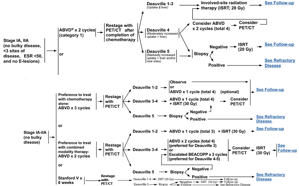
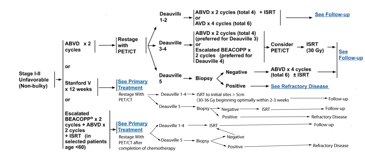
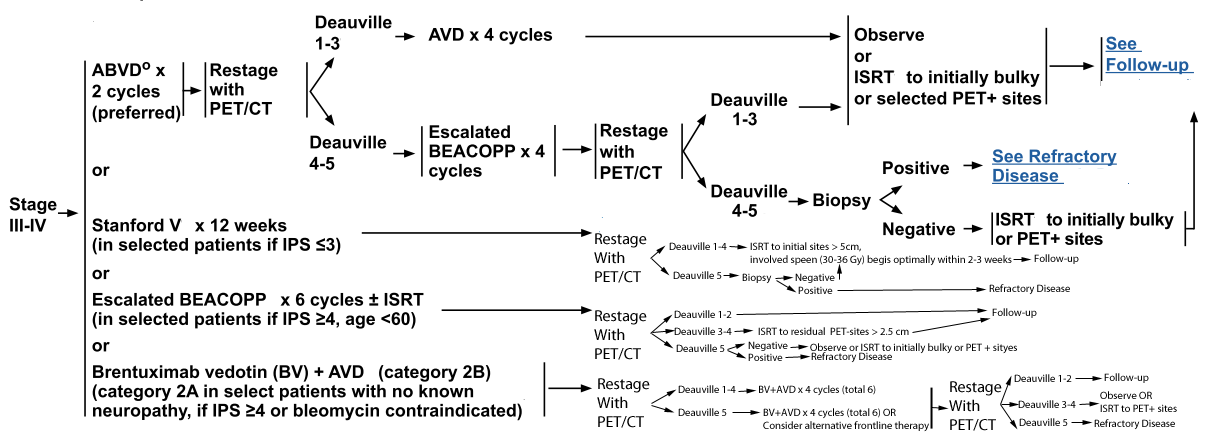
PET 5-POINT SCALE (DEAUVILLE CRITERIA)
Score --- PET/CT scan result
1 ------- No uptake
2 ------- Uptake ≤ mediastinum
3 ------- Uptake > mediastinum but ≤ liver
4 ------- Uptake moderately higher than liver
5 ------- Uptake markedly higher than liver and/or new lesions
X ------- New areas of uptake unlikely to be related to lymphoma
Primary Treatment for Stage IA and IIA, Favorable Classical Hodgkin Lymphoma
Primary Treatment for Stage I and II, Unfavorable Classical Hodgkin Lymphoma

Primary Treatment for Stage III and IV Classical Hodgkin Lymphoma
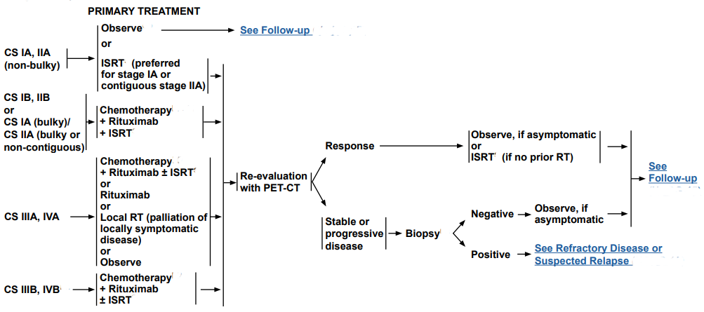
Primary Treatment for Nodular Lymphocyte-Predominant Hodgkin Lymphoma
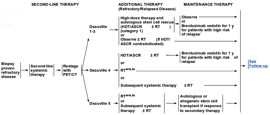
Treatment for Refractory Disease Classical Hodgkin Lymphoma
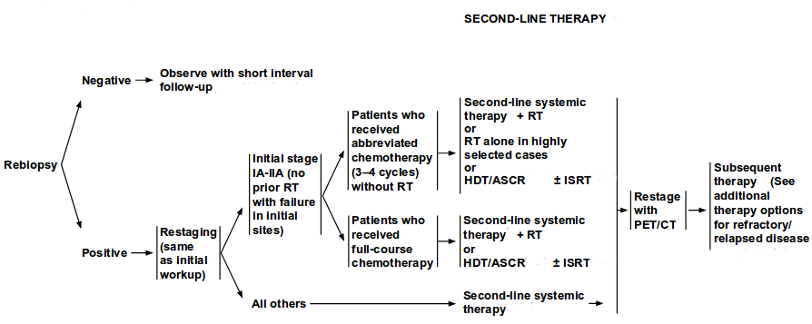
Treatment for Suspected Relapse Classical Hodgkin Lymphoma
Treatment for Refractory or Suspected Relapse Nodular Lymphocyte-Predominant Hodgkin Lymphoma
Treatment for Refractory or Relapsed Disease
NCCN Recommendations for Refractory Disease
Individualized treatment is recommended since there are no data to support a superior outcome with any of the treatment modalities.
Histologic confirmation with biopsy is recommended before initiating treatment for refractory disease.
Although further cytoreduction and HDT/ASCR (with RT if not previously given) are often appropriate, occasional clinical circumstances may warrant the use of RT or
systemic therapy with or without RT.
Conventional-dose second-line systemic therapy may precede HDT/ASCR. RT is recommended when
the sites of relapse have not been previously irradiated. In radiation-naïve patients, TLI may be an appropriate component of HDT/ASCR (high-dose chemoradiotherapy (HDT)/autologous stem-cell rescue (ASCR)).
Second-line systemic therapy followed by response assessment with
PET is recommended for all patients.
Patients with a Deauville score of 1 to 3 should be treated with HDT/ASCR with or without RT or observation with or without RT, if HDT/ASCR is contraindicated.
Additional second-line therapy (RT or second-line systemic therapy with or without RT) is recommended for patients with a Deauville score of 4 or 5.
Alternatively, those with a Deauville score of 4 can be treated with HDT/ASCR with or without RT.
Among patients with relapsed or refractory disease, those with a CR to second-line therapy prior to HDT/ASCR have better outcomes following HDT/ASCR compared to
those with resistant disease.
Everolimus and brentuximab vedotin are included as options for second-line systemic therapy for patients with relapsed or refractory CHL.
Bendamustine and lenalidomide are included as options for additional therapy for patients with relapsed or refractory CHL.
Nivolumab and pembrolizumab are included as additional therapy options for CHL patients that have relapsed or progressed following HDT/ASCR and post-transplant brentuximab vedotin.
The use of brentuximab vedotin as consolidation therapy following HDT/ASCR was evaluated in the AETHERA trial.
In this trial, 329 patients who were at high risk of progression (patients with disease refractory to front-line therapy, relapsed disease <12 months after
frontline therapy, and relapsed disease ≥12 months after frontline therapy with extranodal disease) were randomized (following HDT/ASCR) to brentuximab vedotin (n = 165) or placebo (n = 164).
Patients were required to have obtained a CR, PR, or stable disease to second-line therapy prior to ASCT.
After a median follow-up of 30 months (range 0–50 months), the primary analysis showed that early consolidation with brentuximab vedotin following HDT/ASCR was
associated with improved PFS and the survival benefit was demonstrated across all risk groups.
The median PFS was 42.9 months in the brentuximab vedotin group and 24.1 months in the placebo group. The estimated 2-year PFS rates by independent review
were 63% and 51%, respectively, for the brentuximab vedotin and placebo arms (P = .0013).
There was no statistically significant difference in OS between the two groups (HR 1.15;P = .6204).
Brentuximab vedotin was also well tolerated. Peripheralsensory neuropathy (56%), upper respiratory tract infection (26%), neutropenia (35%), and fatigue (24%) were the most common adverse events.
Based on the results of this study, the panel has included maintenance
therapy with brentuximab vedotin (for one year) following HDT/ASCR
for patients with primary refractory disease. However, the value of this
approach in patients who have received prior treatment with
brentuximab vedotin is not known
and it does not provide a survival benefit.
Allogeneic HSCT with myeloablative conditioning has been associated with lower relapse rate in patients with relapsed or refractory disease;
however, TRM was >50%. Allogeneic HSCT with reduced-intensity conditioning has been reported to have decreased rates of TRM.
However, this approach remains investigational. The panel has included allogeneic HSCT with a category 3 recommendation for select
patients with refractory or relapsed disease.
Conclusion: NCCN Recommendations for R/R Classic HL
Histologic confirmation with biopsy is recommended before initiating treatment for refractory disease.
Second-line systemic therapy is recommended for all patients with refractory disease, followed by response assessment with PET.
Patients with a Deauville score of 1 to 3 should then be treated with HDT/ASCR with or without RT (category 1 recommendation), followed by observation or 1 year of BV maintenance therapy for patients with a high risk of relapse as defined by the AETHERA trial.
RT should be strongly considered for selected sites of relapse that have not been previously irradiated.
If HDT/ASCR is contraindicated, observation with or without RT is recommended.
For patients with a Deauville score of 4 or 5 after second-line systemic therapy, subsequent systemic therapy (±RT) or RT alone is recommended, followed by repeat response assessment. Alternatively, those with a Deauville score of 4 can be treated with HDT/ASCR with or without RT, followed by observation or 1 year of BV maintenance therapy for patients with a high risk of relapse. If patients with a Deauville score of 5 are responsive to secondary therapy, autologous or allogeneic SCT is an option.
NCCN Recommendations for Relapsed Disease
While second-line systemic therapy is an appropriate treatment for any
patient with relapsed disease, regardless of the length of initial
remission, some studies have also suggested that it may not be
essential before proceeding to HDT/ASCR for patients with minimal
residual disease at relapse.
In selected patients with long disease-free intervals and other favorable features, the selection of second-line therapy should be individualized.
Suspected relapse should be confirmed with biopsy. Observation (with short-interval follow-up with PET/CT) is appropriate if biopsy is negative.
Restaging is recommended for patients with positive biopsy.
Second-line systemic therapy with or without ISRT or HDT/ASCR is the
preferred treatment option for patients with stage IA to IIA disease who
were initially treated with chemotherapy alone and experienced failure
at the initial sites. RT alone (conventional or extended field treatment)
may be appropriate for selected patients. All other patients experiencing
disease relapse after initial treatment with chemotherapy or combined
modality therapy should be treated with second-line systemic therapy.
Restaging after completion of treatment is recommended for all
patients. Additional treatment options (based on the score on interim PET scan) are as described for patients with refractory disease.
NCCN Recommendations for the Management of Relapsed or Refractory Disease in Older Adults (Age >60 years)
Outcomes are uniformly poor for elderly patients with relapsed or refractory disease. No uniform recommendation can be made, although clinical trials or possibly single-agent therapy with palliative approach is recommended.
Palliative therapy options include bendamustine, brentuximab vedotin, everolimus,lenalidomide, nivolumab, and pembrolizumab.
Nivolumab and pembrolizumab should be used for patients previously treated with brentuximab vedotin. ISRT alone is an option when systemic therapy is not considered feasible or safe.
Nodular Lymphocyte-Predominant Hodgkin Lymphoma
Patients with refractory or relapsed NLPHL can be managed with second-line therapy as described below. However, some patients have
a chronic indolent disease and may not require aggressive treatment.
Individualized treatment is recommended since there are no data
available to support a superior outcome with any of the treatment
modalities. Rituximab should be considered with all second-
line chemotherapy regimens for patients with relapsed or refractory NLPHL.
NCCN Recommendations
Late relapse or transformation to DLBCL has been reported in patients with NLPHL. In a study of 95 patients diagnosed with NLPHL, with
a median follow-up of 6.5 years, transformation to aggressive lymphoma was seen in 13 (14%) patients and the actuarial risk at 10
and 20 years was 7% and 30%, respectively.
Re-biopsy should be considered to rule out transformation to aggressive
lymphoma prior to initiation of treatment for refractory disease or
suspected disease relapse. Patients with a negative biopsy can be observed.
All patients with biopsy-proven relapsed NLPHL should be observed or treated with second-line therapy (rituximab with or without
chemotherapy or ISRT) followed by reevaluation with PET. No further
treatment is necessary for patients with clinical response. Biopsy is
recommended for patients with progressive disease to rule out
transformation. At this stage, patients should be managed as described
for refractory disease or treated with second-line therapy (rituximab with or without chemotherapy or ISRT) followed by reevaluation with PET.
Maintenance rituximab for 2 years may be considered for patients treated with rituximab alone. Patients with disease transformation to
DLBCL should be managed as discussed in the NCCN Guidelines for Non-Hodgkin’s Lymphomas.
Conclusion: NCCN Recommendations for R/R Classic HL
Suspected relapse at any point should be confirmed with a biopsy.
Observation (with short-interval follow-up with PET/CT) is appropriate if biopsy is negative.
Restaging is recommended for patients with positive biopsy.
Second-line systemic therapy with RT or HDT/ASCR with or without RT are treatment options for patients with stage IA–IIA disease who were initially treated with chemotherapy alone and experienced failure at the initial sites. RT alone may be appropriate in highly selected patients who received abbreviated chemotherapy (3–4 cycles) without RT.
All other patients experiencing disease relapse after initial treatment with chemotherapy or combined modality therapy should be treated with second-line systemic therapy.
Restaging after treatment completion is recommended for all patients. Subsequent treatment options (based on the score on interim PET scan) are described for patients with refractory disease.
PRINCIPLES OF SYSTEMIC THERAPY FOR RELAPSED OR REFRACTORY DISEASE
Regimens
Second-Line Systemic Therapy Options (listed in alphabetical order)
Classical Hodgkin Lymphoma (CHL):
• Brentuximab vedotin alone or in combination with the second-line regimens below
• DHAP (dexamethasone, cisplatin, high-dose cytarabine)
• ESHAP (etoposide, methylprednisolone, high-dose cytarabine, cisplatin)
• Gemcitabine/bendamustine/vinorelbine
• GVD (gemcitabine, vinorelbine, liposomal doxorubicin)
• ICE (ifosfamide, carboplatin, etoposide)
• IGEV (ifosfamide, gemcitabine, vinorelbine)
Nodular Lymphocyte-Predominant Hodgkin Lymphoma (NLPHL):
• DHAP (dexamethasone, cisplatin, high-dose cytarabine)
• ESHAP (etoposide, methylprednisolone, high-dose cytarabine, cisplatin)
• ICE (ifosfamide, carboplatin, etoposide)
• IGEV (ifosfamide, gemcitabine, vinorelbine)
Subsequent Systemic Therapy Options (only for CHL) (listed in alphabetical order):
• Bendamustine
• C-MOPP (cyclophosphamide, vincristine, procarbazine, prednisone) (category 2B)
• Everolimus
• GCD (gemcitabine, carboplatin, dexamethasone)
• Lenalidomide
• MINE (etoposide, ifosfamide, mesna, mitoxantrone)
• Mini-BEAM (carmustine, cytarabine, etoposide, melphalan)
• Nivolumab (For indications, see Checkpoint Inhibitors
• Pembrolizumab (For indications, see Checkpoint Inhibitors
CHECKPOINT INHIBITORS (CPI)
FDA Approved Indications:
• Nivolumab is indicated for the treatment of adult patients with CHL that has relapsed or progressed after:
* Autologous HSCT and brentuximab vedotin; or
* 3 or more lines of systemic therapy that includes autologous HSCT.
• Pembrolizumab is indicated for the treatment of adult and pediatric patients with refractory CHL, or those who have relapsed after 3 or more prior lines of therapy.
General guidelines for use:
• CPI are commonly recommended for:
* Patients with refractory CHL who are transplant-ineligible based on comorbidity or failure of first salvage chemotherapy.
* Any patient who has relapsed after autologous HSCT ± brentuximab vedotin.
• Post-allogeneic transplant, patients can receive either nivolumab or pembrolizumab. There are limited data regarding the use of CPI following allogeneic transplantation. Caution is advised due to increased risk of GVHD (graft-versus-host disease) and other immunological complications.
Updates to the Treatment of R/R Classic HL
Brentuximab Vedotin
BV, a CD30-directed antibody-drug conjugate, has demonstrated activity in patients with R/R CD30+ lymphomas. In a pivotal phase II multicenter study of 102 patients with R/R HL after HDT/ASCR, BV induced objective responses and complete remissions in 75% and 34% of patients, respectively, with a median follow-up of >1.5 years. Median PFS for all patients and the median duration of response for those in complete remission were 5.6 and 20.5 months, respectively.17 The 3-year follow-up data confirmed durable remissions in patients whose disease responded to BV.15 After a median follow-up of approximately 3 years, the estimated median OS and PFS were 40.5 and 9.3 months, respectively. In those who experienced a complete remission on BV, the estimated 3-year OS and PFS rates were 73% and 58%, respectively.
Recent studies are investigating the utility of BV as second-line therapy for R/R HL, either sequentially or in combination with other regimens, prior to HDT/ASCR.11,52–56 A multicenter prospective phase II study examined the efficacy of BV in patients (n=37) with R/R HL (prior to HDT/ASCR), which resulted in an ORR of 68% (CR, 35%), and 33 patients (89%) successfully proceeded to HDT/ASCR.52 Other studies have combined BV with bendamustine, ICE (ifosfamide/carboplatin/etoposide), or ESHAP (etoposide/methylprednisolone/high-dose cytarabine/cisplatin), with preliminary data demonstrating PET-negative responses ranging from approximately 75% to 90%.11,53,54,56 During the 2018 guideline update, the NCCN panel included a recommendation that BV could be used alone or in combination with second-line systemic therapy options for R/R HL.
Use of BV as consolidation or maintenance therapy following HDT/ASCR was evaluated in the AETHERA trial. In this trial, 329 patients who were at high risk of progression after HDT/ASCR (patients with disease refractory to frontline therapy, relapsed disease <12 months after frontline therapy, or extranodal disease at the start of pretransplantation salvage chemotherapy) were randomized to BV (n=165) or placebo (n=164).57 Patients were required to have obtained a complete remission, partial remission, or stable disease to second-line therapy prior to ASCT. After a median follow-up of 30 months (range, 0–50 months), primary analysis showed that early consolidation with BV after HDT/ASCR was associated with improved PFS, and the survival benefit was demonstrated across all risk groups. Median PFS was 42.9 months in the BV group and 24.1 months in the placebo group. The estimated 2-year PFS rates by independent review were 63% and 51%, respectively, for the BV and placebo arms (P=.0013). The benefit of BV on PFS in this context appeared to decrease in patients who were PET-negative before HDT/ASCR.
During the 2018 update, the panel clarified the patient subpopulation likely to gain maximal benefit from BV use as maintenance therapy for 1 year after HDT/ASCR. These are patients with high risk of relapse with ≥2 of the following risk factors: remission duration of <1 year, extranodal involvement, PET-positive at transplant, B symptoms, and/or >1 salvage/subsequent therapy regimen.
Programmed Death Receptor-1 Inhibitors
Blockade of the programmed death receptor-1 (PD-1), an immune checkpoint, has demonstrated activity in patients with R/R PD-1–positive lymphomas.12–14,18,37,58 In a phase I study of 23 patients with R/R HL pretreated with both HDT/ASCR and BV, treatment with nivolumab, a PD-1 inhibitor, induced an ORR of 87%, with a PFS rate of 86% at 24 weeks.12 In a phase II study of 80 patients with R/R HL pretreated with both HDT/ASCR and BV, treatment with nivolumab induced an objective response in 53 of 80 patients (66.3%; 95% CI, 54.8–76.4), as determined by an independent radiologic review committee, at a median follow-up of 8.9 months.18 In a phase I study of 31 patients with R/R HL pretreated with BV, treatment with pembrolizumab, another PD-1 inhibitor, induced a complete remission rate of 16% (90% CI, 7%–31%) and a partial remission rate of 48% resulting in an ORR of 65% (90% CI, 48%–79%).13 In a phase II study of 210 patients with R/R HL, the efficacy of pembrolizumab was examined in 3 cohorts of patients with disease progression after (1) ASCT and subsequent BV, (2) salvage chemotherapy and BV (ineligible for ASCT due to chemoresistant disease), and (3) ASCT without BV14; the corresponding ORRs were 73.9%, 64.2%, and 70%, respectively.14 During the 2018 guideline update, the panel clarified and expanded on contexts for the use of the PD-1 inhibitors nivolumab and pembrolizumab as subsequent systemic therapy options for R/R classic HL (see HODG-E 1 and 2 of 3; pages 249 and 250, respectively). Emerging data are investigating the combination of BV and checkpoint inhibitors as an option for R/R HL prior to transplant.
Summary
HL is an uncommon malignancy involving lymph nodes and the lymphatic system. CHL and NLPHL are the two main types of HL. CHL is characterized by the presence of Reed-Sternberg cells in an inflammatory background, whereas NLPHL is characterized by the presence of lymphocytic and histiocytic cells.
Current management of CHL involves initial treatment with chemotherapy or combined modality therapy, followed by restaging with
PET/CT to assess treatment response using the Deauville criteria (5-PS).
Combined modality therapy (ABVD, ABVD followed by escalated BEACOPP or Stanford V plus ISRT) or ABVD alone are included as treatment options for patients with stage IA or IIA favorable CHL.
Chemotherapy (ABVD or Stanford V or BEACOPP plus ABVD) followed by consolidative ISRT is recommended for patients with stage I-II unfavorable disease.
Chemotherapy with ABVD or Stanford V or escalated-dose BEACOPP is recommended for patients with stage III -IV disease.
HDT/ASCR is the best treatment option for patients with refractory or relapsed CHL, although it does not improve OS.
Second-line therapy (RT or second-line systemic therapy with or without RT) may be given prior to HDT/ASCR.
Maintenance therapy with brentuximab vedotin (for one year) following HDT/ASCR is included as an option for patients with primary refractory disease.
ISRT is the preferred treatment for patients with stage IA or IIA non-bulky NLPHL.
Observation may be an option for highly selected patients with stage IA disease with a completely excised solitary node.
A brief course of chemotherapy plus ISRT with rituximab is recommended for patients with stage IB or IIB disease and for very rare patients presenting with stage IA or IIA bulky disease.
Chemotherapy with rituximab with or without ISRT is recommended for all patients with stage III-IV disease.
Alternatively, selected patients with stage IIIA-IVA disease can either be observed or treated with local palliative RT or rituximab.
Late relapse or transformation to DLBCL has been reported in patients with NLPHL. In patients with suspected relapse, re-biopsy should be considered to rule out transformation to DLBCL.
Patients with refractory or relapsed NLPHL can be managed with second-line therapy. However, some patients have a chronic indolent disease and may not require aggressive treatment, unless they are symptomatic.
HL is now curable in most patients because of the introduction of more effective and less toxic regimens. However, survivors may experience
late treatment-related side effects. For this reason, long-term follow-up by an oncologist is essential after completion of treatment. Counseling about issues of survivorship and careful monitoring for late treatment-related side effects should be an integral part of follow-up.
Consistent with NCCN philosophy, participation in clinical trials is always encouraged.