Multiple Myeloma and Plasma Dyscrasia
Plasma Cell Neoplasms
There are several types of plasma cell neoplasms. These diseases are all associated with a monoclonal (or myeloma) protein (M protein). They include monoclonal gammopathy of undetermined significance (MGUS), isolated plasmacytoma of the bone, extramedullary plasmacytoma, and multiple myeloma.
Incidence and Mortality
Estimated new cases and deaths from multiple myeloma in the United States in 2018:
•New cases: 30,770. •Deaths: 12,770.
Introduction
Myeloma is a blood cancer of cells found in the bone marrow, specifically the so-called “plasma cells.” The bone marrow is the spongy tissue inside your bones that normally generates the components of your blood. Myeloma begins when healthy plasma cells change and grow out of control. Plasma cells are normally a key part of the body's immune system. In particular, they produce antibodies that help the body fight infection. Abnormal plasma cells can crowd out or suppress the growth of other cells in the bone marrow that produce red blood cells, white blood cells, and platelets. They also reduce the creation of normal plasma cells, which lowers a person’s immunity. This suppression may result in:
• Anemia, from a shortage of red blood cells
• Excessive bleeding from cuts, from a shortage of platelets
• Decreased ability to fight infection, from a shortage of white blood cells and the body’s inability to respond to infection because of the presence of abnormal antibodies
It is important to explain that like regular plasma cells, myeloma cells can produce antibodies. However, myeloma cells are unable to produce healthy, functioning antibodies. Instead, they make what is called “monoclonal protein,” or “M protein.” M protein can build up in the blood and urine, potentially damaging the kidneys and other organs, as well as reducing immunity. A healthy person who is found to have a small amount of this M protein is said to have monoclonal gammopathy of undetermined significance (MGUS).
Myeloma causes structural bone damage, which can result in weakened bones and leads to painful fractures or bone breaks over time. Myeloma is usually called multiple myeloma because most people (90% or more) have multiple bone lesions when diagnosed or lesions develop over the course of the illness.
Solitary plasmacytoma is a mass of myeloma cells that involves only 1 site in the bone or, less commonly, other organs, such as in the upper respiratory tract, including the nose and throat or gastrointestinal systems.
Extramedullary plasmacytoma describes myeloma that started outside the bone marrow in locations such as the lymph glands, sinuses, throat, liver, digestive tract, or under the skin.
Risk Factors
The causes of myeloma are not known or well understood. There are currently no known ways to prevent it. Currently, there are also no obvious strong risk factors for myeloma. Mutations or changes in plasma cells are acquired, not inherited, so having a relative with the disease usually does not mean another family member is at higher risk for developing it. There appears to be a slight increase in the incidence of the disease in first-degree relatives (parents or siblings) of people with multiple myeloma.
The following factors can raise a person's risk of developing myeloma:
• Age. Myeloma occurs most commonly in people over 60. The average age at diagnosis is 70. Only 2% of cases occur in people under 40.
• Race. Myeloma occurs twice as frequently in black people than in white people. The reasons why are unclear, although it appears to also be more common in the Middle East, North Africa, and the Mediterranean.
• Exposure to radiation or chemicals. People who have been exposed to radiation or to asbestos, benzene, pesticides, and other chemicals used in rubber manufacturing may be at higher risk for developing myeloma. Those exposed to wood products, such as carpenters, furniture makers, and paper makers, are also at higher risk. There is also a high incidence of myeloma among professional firefighters and those exposed to herbicides, including Agent Orange.
• Personal history. People with a history of a solitary plasmacytoma of the bone are at greater risk for developing multiple myeloma.
• Monoclonal gammopathy of undetermined significance (MGUS). As explained in the Introduction, a person with a small amount of M protein in his or her blood has a 1% to 2% chance of developing myeloma, lymphoma, or another blood-related cancer called Waldenstrom’s macroglobulinemia per year (see the Stages section for more information).
• Gender. Myeloma is slightly more common in men.
Clinical Presentation and Evaluation
| Plasma Cell Neoplasm | M Protein Type | Pathology | Clinical Presentation |
| MGUS (Monoclonal Gammopathy of Undetermined Significance) | IgG kappa or lambda; or IgA kappa or lambda | <10% plasma cells in bone marrow | Asymptomatic, with minimal evidence of disease (aside from the presence of an M protein) |
| Isolated plasmacytoma of bone | IgG kappa or lambda; or IgA kappa or gamma | Solitary lesion of bone; <10% plasma cells in marrow of uninvolved site | Asymptomatic or symptomatic |
| Extramedullary plasmacytoma | IgG kappa or lambda; or IgA kappa or gamma | Solitary lesion of soft tissue; most commonly occurs in the nasopharynx, tonsils, or paranasal sinuses | Asymptomatic or symptomatic |
| Multiple myeloma | IgG kappa or lambda; or IgA kappa or gamma | Often, multiple lesions of bone | Symptomatic |
Evaluation of patients with monoclonal (or myeloma) protein (M protein)
Idiotypic myeloma cells can be found in the blood of myeloma patients in all stages of the disease. For this reason, when treatment is indicated, systemic treatment must be considered for all patients with symptomatic plasma cell neoplasms. Patients with MGUS or asymptomatic, smoldering myeloma do not require immediate treatment but must be followed carefully for signs of disease progression.
The major challenge is to separate the stable, asymptomatic group of patients who do not require treatment from patients with progressive, symptomatic myeloma who may need to be treated immediately.
Patients with a monoclonal (or myeloma) protein (M protein) in the serum and/or urine are evaluated by some of the following criteria:
• Measure and follow the serum M protein by serum electrophoresis or by specific immunoglobulin (Ig) assays; however, specific Ig quantification always overestimates the M protein because normal Ig are included in the result. For this reason, the preference is often that baseline and follow-up measurements of the M protein be done by the same method. Quantitative serum-free light chains (FLC) may be helpful to follow response if an M protein is not apparent.
• Measure and follow the amount of M-protein light chains excreted in the urine over 24 hours. Measure the total amount of protein excreted over 24 hours and multiply this value by the percentage of urine protein that is M protein, as determined by electrophoresis of concentrated urine protein. An easier, but less accurate, method uses a spot-urine protein electrophoresis.
• Identify the heavy and light chain of the M protein by immunofixation electrophoresis.
• Measure the hemoglobin, leukocyte, platelet, and differential counts.
• Determine the percentage of marrow plasma cells. Be aware that marrow plasma-cell distribution may vary in different sites. Bone marrow is often sent for cytogenetics and fluorescence in situ hybridization testing for genetic markers of high-risk disease.
• Measure serum-free kappa and lambda light chain. This is especially useful in cases of oligosecretory plasma-cell dyscrasia or for following cases of light-chain amyloidosis. The FLC ratio of over 100 can predict a greater than 70% progression within 2 years in patients with smoldering myeloma.
• If clinically warranted, take needle aspirates of a solitary lytic bone lesion, extramedullary tumor(s), or enlarged lymph node(s) to determine whether these are plasmacytomas.
• Evaluate renal function with serum creatinine and a creatinine clearance.
• Electrophoresis of concentrated urine protein is very helpful in differentiating glomerular lesions from tubular lesions. Glomerular lesions, such as those resulting from glomerular deposits of amyloid or light-chain deposition disease, result in the nonselective leakage of all serum proteins into the urine; the electrophoresis pattern of this urine resembles the serum pattern with a preponderance of albumin.
In most myeloma patients, the glomeruli function normally allows only the small molecular weight proteins, such as light chains, to filter into the urine. The concentration of protein in the tubules increases as water is reabsorbed. This leads to precipitation of proteins and the formation of tubular casts, which may injure the tubular cells. With tubular lesions, the typical electrophoresis pattern shows a small albumin peak and a larger light-chain peak in the globulin region; this tubular pattern is the usual pattern found in myeloma patients.
• Measure serum levels of calcium, alkaline phosphatase, lactic dehydrogenase, and, when indicated by clinical symptoms, cryoglobulins and serum viscosity.
• Obtain radiographs of the skull, ribs, vertebrae, pelvis, shoulder girdle, and long bones.
• Obtain a spinal magnetic resonance imaging (MRI) scan (or spinal computed tomography [CT] or positron emission tomography (PET)–CT scan depending on availability) if the skeletal survey is negative. At diagnosis, whole-body PET scan or MRI of the total spine and pelvis appears equally efficacious in the detection of bone lesions.
• If amyloidosis is suspected, perform a needle aspiration of subcutaneous abdominal fat and stain the bone marrow biopsy for amyloid as the easiest and safest way to confirm the diagnosis.
• Measure serum albumin and beta-2-microglobulin as independent prognostic factors.
• The presence of circulating myeloma cells is considered a poor prognostic factor. Primary plasma cell leukemia has a particularly poor prognosis.
These initial studies are often compared with subsequent values at a later time, when it is necessary to decide whether the disease is stable or progressive, responding to treatment, or getting worse.
As mentioned before, the major challenge is to separate the stable, asymptomatic group of patients who do not require treatment from patients with progressive, symptomatic myeloma who may need to be treated immediately.
It is important to note that the intravenous contrast dye often used for CT scans for other types of cancer is specifically avoided in people with multiple myeloma. Tell the radiologist or the radiology technician about your diagnosis before receiving dye injection into your vein, because this can cause kidney damage in people with myeloma. (ASCO)
Monoclonal Gammopathy of Undetermined Significance (MGUS)
Patients with MGUS have an M protein in the serum without findings of multiple myeloma, macroglobulinemia, amyloidosis, or lymphoma and have fewer than 10% of plasma cells in the bone marrow. Patients with smoldering myeloma have similar characteristics but may have more than 10% of plasma cells in the bone marrow.
These types of patients are asymptomatic and do not need to be treated. Patients with MGUS and risk factors for disease progression, however, must be followed carefully because they are more likely to develop myeloma (most commonly), amyloidosis, lymphoplasmacytic lymphoma, or chronic lymphocytic leukemia and may then require therapy.
Virtually all cases of multiple myeloma are preceded by a gradually rising level of MGUS. The annual risk of progression of MGUS to a lymphoid or plasma cell malignancy ranges from 0.5% to 1.0% in population-based cohorts. This risk ranges from 2% to more than 20% in higher-risk patients.
Risk factors that predict disease progression include the following:
• An abnormal serum-FLC ratio.
• Non-IgG class MGUS.
• A high serum-M protein level (≥15 g/L).
A Swedish cohort study confirmed the higher-risk factors of abnormal serum-FLC ratio and the high serum–monoclonal protein level. They described the additional risk factor of immunoparesis, which is defined as the reciprocal depression of the other Ig classes (if a patient has an IgG kappa M-protein, the IgM and IgA would be below normal levels with immunoparesis). Incorporation of gene-expression profiles to better assess risk is also under clinical evaluation.
Monoclonal gammopathies that cause organ damage, particularly to the kidney, heart, or peripheral nerves require immediate therapy with the same strategies applied for the conventional plasma-cell dyscrasias. A monoclonal gammopathy causing renal dysfunction—by direct antibody deposition or amyloidosis—is referred to as monoclonal gammopathy of renal significance. Rising serum creatinine, dropping glomerular filtration rates, and increasing urinary–albumin excretion are all parameters that may signify renal damage and are assessed prospectively for high-risk MGUS patients. Although the N-terminal pro-brain natriuretic peptide is a very sensitive marker for amyloid involvement in the heart, the low specificity must be noted. These extra tests are included with the M-protein level, FLC levels, and FLC ratio when following patients with MGUS.
Isolated Plasmacytoma of Bone
The patient has an isolated plasmacytoma of the bone if the following are found:
• A solitary lytic lesion of plasma cells on skeletal survey in an otherwise asymptomatic patient.
• A bone marrow examination from an uninvolved site contains less than 10% plasma cells. The absence of plasma cells on flow cytometry of the bone marrow suggests a low (<10%) risk of recurrence after radiation therapy of the isolated bone plasmacytoma.
MRI may reveal unsuspected bony lesions that were undetected on standard radiographs. MRI scans of the total spine and pelvis may identify other bony lesions.
Extramedullary Plasmacytoma
A patient has extramedullary plasmacytoma if the following are found:
• Isolated plasma-cell tumors of soft tissues, most commonly occurring in the tonsils, nasopharynx, or paranasal sinuses.
• Negative findings on skeletal x-rays and bone marrow biopsy.
Multiple Myeloma
Multiple myeloma is a systemic malignancy of plasma cells that typically involves multiple sites within the bone marrow and secretes all or part of a monoclonal antibody.
Prognosis
Multiple myeloma is highly treatable but rarely curable. The median survival in the prechemotherapy era was about 7 months. After the introduction of chemotherapy, prognosis improved significantly with a median survival of 24 to 30 months and a 10-year survival rate of 3%. Even further improvements in prognosis have occurred because of the introduction of newer therapies such as pulse corticosteroids, thalidomide, lenalidomide, bortezomib, and autologous and allogeneic stem cell transplantation, with median survivals now exceeding 45 to 60 months. Patients with plasma cell leukemia or with soft tissue plasmacytomas (often with plasmablastic morphology) in association with multiple myeloma have poor outcomes.
Multiple myeloma is potentially curable when it presents as a solitary plasmacytoma of bone or as an extramedullary plasmacytoma. (Refer to the Isolated Plasmacytoma of Bone and Extramedullary Plasmacytoma sections of this summary for more information.)
Amyloidosis Associated With Plasma Cell Neoplasms
Multiple myeloma and other plasma cell neoplasms may cause a condition called amyloidosis. Primary amyloidosis can result in severe organ dysfunction especially in the kidney, heart, or peripheral nerves. Clinical symptoms and signs include the following:
• Fatigue.
• Purpura.
• Enlarged tongue.
• Diarrhea.
• Edema.
• Lower-extremity paresthesias.
Accurate diagnosis of amyloidosis requires histologic evidence of amyloid deposits and characterization of the amyloidogenic protein using immunoelectron microscopy. In one series of 745 consecutive patients, 20% of patients with non-amyloid light chain amyloidosis (usually transthyretin) had an innocent monoclonal gammopathy, indicating the significant risk of misdiagnosis.
Elevated serum levels of cardiac troponins, amino-terminal fragment brain-type natriuretic peptide, and serum-FLC are poor prognostic factors.[48,49] A proposed staging system for primary systemic amyloidosis based on these serum levels requires independent and prospective confirmation. An increase in levels of serum-FLC over many years can precede the clinical diagnosis of amyloid light-chain (AL) amyloidosis.
POEMS Syndrome
POEMS (polyneuropathy, organomegaly, endocrinopathy, monoclonal gammopathy, and skin changes) syndrome is a rare paraneoplastic condition associated with a plasma cell dyscrasia of early or late stage. The acronym describes a constellation of findings often marked by polyneuropathy, organomegaly (usually splenomegaly), endocrinopathy, monoclonal plasma cell dyscrasia, and skin changes. Both sclerotic or lytic bone lesions and lymphadenopathy (with possible Castleman's histology) may be identified. Anecdotal reports suggest remissions using myeloma-directed therapy.
Stage Information About Plasma Cell Neoplasms
No generally accepted staging system exists for monoclonal gammopathy of undetermined significance (MGUS), isolated plasmacytoma of bone, or extramedullary plasmacytoma. Of the plasma cell neoplasms, a staging system exists only for multiple myeloma.
Multiple Myeloma
Multiple myeloma is staged by estimating the myeloma tumor cell mass on the basis of the amount of monoclonal (or myeloma) protein (M protein) in the serum and/or urine, along with various clinical parameters, such as hemoglobin and serum calcium concentrations, the number of lytic bone lesions, and the presence or absence of renal failure. Impaired renal function worsens prognosis regardless of stage.
The stage of the disease at presentation is a strong determinant of survival, but it has little influence on the choice of therapy since almost all patients, except for rare patients with solitary bone tumors or extramedullary plasmacytomas, have generalized disease.
International staging system
The International Myeloma Working Group (IMWG) studied 11,171 patients, of whom 2,901 received high-dose therapy and 8,270 received only standard-dose therapy. The IMWG evaluated 4,445 patients to create a Revised International Staging System (R-ISS) incorporating lactate dehydrogenase (LDH) levels and interphase fluorescence in situ hybridization (I-FISH) results.
An International Staging System (ISS) was derived and is shown below in Table.
| Stage | Criteria | Median Survival (mo) |
| I | Beta-2-microglobulin <3.5 mg/L and albumin ≥3.5 g/dL | Not reached |
| II | Not R-ISS I or III | 83 |
| III | Beta-2-microglobulin ≥5.5 mg/L and either high LDH or high-risk chromosomal abnormalities by I-FISH (defined as presence of del (17p) and/or translocation t(4;14) and/or translocation t(14;16)) | 43 |
I-FISH = interphase fluorescence in situ hybridization; R-ISS = Revised International Staging System.
Genetic factors and risk groups
Newer clinical investigations are stratifying patients with multiple myeloma into so-called good-risk, intermediate-risk, and high-risk groups, based on genetic aberrations detected by I-FISH. (See Table below.) This stratification, based on cytogenetic findings, has been derived from retrospective analyses and requires prospective validation. Bone marrow samples are sent for cytogenetic and FISH analysis. Plasma cell leukemia has a particularly poor prognosis. The otherwise favorable prognosis of hyperploidy is trumped by coexistent adverse cytogenetics.
| Risk Group | Cytogenetic Findings | Disease Characteristics | Median Survival (y) |
| Good risk | Has any of the following cytogenetic findings: (1) no adverse FISH or cytogenetics, (2) hyperdiploidy, (3) t(11;14) by FISH, or (4) t(6;14) by FISH. | These patients most often have (1) disease that expresses IgG kappa monoclonal gammopathies and (2) lytic bone lesions. | 8–10 |
| Intermediate risk | t(4;14) by FISH | These patients often have IgA lambda monoclonal gammopathies and less bone disease. | 5 |
| High risk | Has any of the following cytogenetic findings: (1) del 17p by FISH, (2) t(14;16) by FISH, (3) t(4;14), (4) t(14;20), (5) cytogenetic del 13, (6) nonhyperdiploidy without adverse cytogenetic findings, (7) 1q gain, or (8) plasma cell leukemia. | These patients have (1) disease that expresses IgA lambda monoclonal gammopathies (often) and (2) skeletal-related complications (less often). | <2 |
Treatment Option Overview for Plasma Cell Neoplasms
The major challenge in treating plasma cell neoplasms is to separate the stable, asymptomatic group of patients who do not require immediate treatment from patients with progressive, symptomatic myeloma who may need to be treated immediately. Monoclonal gammopathy of undetermined significance or smoldering myeloma must be distinguished from progressive myeloma.
Asymptomatic Plasma Cell Neoplasms (Smoldering Multiple Myeloma)
Asymptomatic patients with multiple myeloma who have no lytic bone lesions and normal renal function may be initially observed safely outside the context of a clinical trial.
Increasing anemia is the most reliable indicator of progression.
The following criteria represent the new definition for smoldering myeloma:
• Serum monoclonal protein immunoglobulin (Ig) G or IgA of at least 30 g/L or urinary monoclonal protein of at least 500 mg per 24 hours.
• Clonal bone marrow plasma cells 10% to 60% (>60% represents overt myeloma).
• Absence of amyloidosis or myeloma-defining events as follows:
• Hypercalcemia greater than 1 mg/dl higher than normal.
• Creatinine greater than 2 mg/dl or creatinine clearance less than 40 ml/min.
• Anemia with hemoglobin less than 10.0 g/dl.
• Bone lesions (one or more) on skeletal radiography, computed tomography (CT) or positron emission tomography (PET)-CT.
• Clonal plasma cell percentage in marrow at 60% or more.
• Involved: uninvolved serum-free light chain (FLC) ratio at 100 or more.
• More than one focal lesion of at least 5 mm on magnetic resonance imaging (MRI) of the spine.
A prospective, randomized clinical trial investigated the role of immediate therapy for patients with smoldering multiple myeloma by specifying high-risk patients with both 10% or more marrow plasma cells and a serum monoclonal (or myeloma) protein (M protein) of at least 3 g/dL. The trial randomly assigned 125 patients to receive lenalidomide plus dexamethasone or observation.
• With a median follow-up of 75 months, lenalidomide plus dexamethasone provided benefit in time to progression compared with observation, with a median time not reached (95% confidence interval [CI], 47 months to not reached) compared with 23 months (95% CI, 16‒31 months) (hazard ratio, 0.24; 95% CI, 0.14‒0.41).[Level of evidence: 1iiDiii]
• There was no difference in overall survival (OS) at a median follow-up of 75 months.
• At the beginning of this trial, some of the patients had what would now be considered overt myeloma, based on the updated criteria listed above. This may influence the interpretation of the study because overt myeloma patients might be responsible for some of the benefits seen with therapy.
lenalidomide plus dexamethasone regimen:
lenalidomide 25 mg/day p.o. days 1-21
dexamethasone 40 mg/day p.o. days 1,8,15,21
Every 28 days for 6 cycles
Symptomatic Plasma Cell Neoplasms
Patients with symptomatic advanced disease require treatment.
Treatment most often is directed at reducing the tumor cell burden and reversing any complications of disease, such as renal failure, infection, hyperviscosity, or hypercalcemia, with appropriate medical management. The International Myeloma Working Group (IMWG) has published new criteria for identifying patients with active myeloma who require therapy. These criteria include the following:
• Amyloidosis.
• Hypercalcemia greater than 1 mg/dl higher than normal.
• Creatinine greater than 2 mg/dl or creatinine clearance less than 40 ml/min. Myeloma can cause renal dysfunction via hypercalcemia, amyloidosis, or light chain deposition disease.
• Anemia with hemoglobin less than 10.0 g/dl.
• Bone lesions (one or more) on skeletal radiography, whole-body MRI or spine and pelvis MRI, or PET-CT scans.
• Clonal plasma cell percentage in marrow at 60% or more.
• Involved: uninvolved serum-FLC ratio at 100 or more.
• More than one focal lesion of at least 5 mm on skeletal bone survey, or if negative, total-body MRI, or MRI of the spine and pelvis, or PET-CT scan.
Response criteria have been developed for patients on clinical trials by the IMWG. A very good partial response (VGPR) is defined as a reduction of 90% or more in the serum monoclonal protein and a 24-hour urine monoclonal protein of less than 100 mg. Although not incorporated in the IMWG criteria, many trials report near complete response (nCR) when patients have less than 5% bone marrow plasma cells and unmeasurable serum monoclonal proteins but still have positive serum and/or urine immunofixation. Note that these nCR patients are incorporated into the VGPR group by the IMWG. Patients who achieve a CR by IMWG criteria (with a negative immunofixation along with the clear marrow and unmeasurable serum monoclonal proteins) are often said to have attained a stringent CR if they also normalize their free kappa/lambda light–chain levels and ratio. The clinical utility of these various categories must be validated in clinical trials. Whether these response definitions will translate into clinically meaningful endpoints, such as OS, remains to be seen.
Current therapy for patients with symptomatic myeloma can be divided into the following categories:
• Induction therapies.
• Consolidation therapies, which are less applicable for the very elderly.
• Maintenance therapies.
• Supportive care, such as bisphosphonates.
Treatment for Amyloidosis Associated With Plasma Cell Neoplasms
Treatment depends on assessing the extent of systemic damage from the amyloidosis and the underlying plasma cell dyscrasia.
A rising and elevated level of N-terminal pro brain natriuretic peptide (NT-proBNP) may predict impending cardiac failure in the setting of cardiac amyloidosis, and early treatment should be considered for these patients.
Treatment Options for Amyloidosis Associated With Plasma Cell Neoplasms
1. Chemotherapy, immunomodulatory (IMiDs) agents, and proteasome inhibitors.
2. Stem cell rescue.
1. Chemotherapy
As is true for all plasma cell dyscrasias, responses have been reported for all the same regimens active in multiple myeloma.
Two randomized trials showed prolonged overall survival (OS) with the use of oral chemotherapy with melphalan with or without colchicine versus colchicine alone.[Level of evidence: 1iiA]
2. Stem cell rescue
A randomized, prospective study of 100 patients with immunoglobulin light-chain amyloidosis compared melphalan plus high-dose dexamethasone with high-dose melphalan plus autologous stem cell rescue.
After a median follow-up of 3 years, median OS favored the nontransplant arm (56.9 months vs. 22.2 months; P = .04).[Level of evidence: 1iiA]
The 24% transplant-related mortality in this series and others reflects the difficulties involved with high-dose chemotherapy in older patients with organ dysfunction.
Between 2007 and 2012, the International Blood and Marrow Transplant Research Program identified 800 patients with amyloidosis who underwent autologous stem cell transplantation; the 5-year OS was 77% and transplant-related mortality was 5%, suggesting better selection of patients for transplantation.[Level of evidence: 3iiiA]
A randomized trial confirming the benefit of autologous transplantation is not anticipated.
An anecdotal series describes full-intensity and reduced-intensity allogeneic stem cell transplantation.
Treatment for Monoclonal Gammopathy of Undetermined Significance
Treatment Options for Monoclonal Gammopathy of Undetermined Significance (MGUS)
Watchful waiting
Multiple myeloma, other plasma cell dyscrasia, or lymphoma will develop in 12% of patients by 10 years, 25% by 20 years, and 30% by 25 years.
All patients with MGUS are generally kept under observation to detect increases in M protein levels and development of a plasma cell dyscrasia. Higher levels of initial M protein levels may correlate with increased risk of progression to multiple myeloma. In a large retrospective report, the risk of progression at 20 years was 14% for an initial monoclonal protein level of 0.5 g/dL or less, 25% for a level of 1.5 g/dL, 41% for a level of 2.0 g/dL, 49% for a level of 2.5 g/dL, and 64% for a level of 3.0 g/dL.
Treatment is delayed until the disease progresses to the stage that symptoms or signs appear.
Patients with MGUS or smoldering myeloma do not respond more frequently, achieve longer remissions, or have improved survival if chemotherapy is started early while they are still asymptomatic as opposed to waiting for progression before treatment is initiated. Newer therapies have not been proven to prevent or delay the progression of MGUS to a plasma cell dyscrasia.
Treatment for Isolated Plasmacytoma of Bone
Treatment Options for Isolated Plasmacytoma of Bone
1. Radiation therapy to the lesion.
2. Chemotherapy (if the monoclonal [or myeloma] protein [M protein] increases and other evidence of symptomatic multiple myeloma occurs).
1. Radiation therapy
About 25% of patients have a serum and/or urine M protein; generally this disappears following adequate radiation therapy to the lytic lesion.
The survival rate of patients with isolated plasmacytoma of bone treated with radiation therapy to the lesion is greater than 50% at 10 years, which is much better than the survival rate of patients with disseminated multiple myeloma.
2. Chemotherapy
Most patients will eventually develop disseminated disease and require chemotherapy; almost 50% of them will do so within 2 years of diagnosis. However, patients with serum paraprotein or Bence Jones protein, who have complete disappearance of these proteins after radiation therapy, may be expected to remain free of disease for prolonged periods. Patients with a negative flow cytometry on bone marrow examination for plasma cell infiltration are also unlikely to relapse. Patients who progress to multiple myeloma tend to have good responses to chemotherapy with a median survival of 63 months after progression.
Treatment for Extramedullary Plasmacytoma
Treatment Options for Extramedullary Plasmacytoma
1. Radiation therapy to the isolated lesion with fields that cover the regional lymph nodes, if possible.
2. In some cases, surgical resection may be considered, but it is usually followed by radiation therapy.
3. If the monoclonal (or myeloma) protein (M protein) persists or reappears, the patient may need further radiation therapy. In some patients, the plasmacytoma may shrink, but not disappear, and the M protein persists. Close follow-up is generally warranted for these types of patients. Surgery often is performed if the plasmacytoma is in a site where it can be removed easily (e.g., in the tonsil); the M protein may disappear from the blood or urine. In other cases, persistence or an increasing M protein may herald progression to multiple myeloma.
4. Chemotherapy is required if the disease progresses and causes symptoms.
Patients with isolated plasma cell tumors of soft tissues, most commonly occurring in the tonsils, nasopharynx, or paranasal sinuses, may need to have skeletal x-rays and bone marrow biopsy (both of which are most often negative) and evaluation for M protein in serum and urine.
About 25% of patients have serum and/or urine M protein; this frequently disappears following adequate radiation.
Extramedullary plasmacytoma is a highly curable disease with progression-free survival ranging from 70% to 87% at 10 to 14 years after treatment with radiation therapy (with or without previous resection).
Treatment for Multiple Myeloma
Initial Evaluation
The initial approach to the patient is to evaluate the following parameters:
1. Detection and quantification of a monoclonal (or myeloma) protein (M protein) in the serum or urine, and possible immunoparesis (suppression of the other uninvolved immunoglobulins).
2. Detection of more than 10% of plasma cells on a bone marrow examination, along with flow cytometry, cytogenetics, and fluorescence in situ hybridization testing.
3. Detection of lytic bone lesions or generalized osteoporosis in skeletal x-rays, or whole-body or spinal and pelvic magnetic resonance imaging scans, or focal bone lesions on positron emission tomography-computed tomography scan.
4. Presence of soft tissue plasmacytomas.
5. Serum albumin and beta-2-microglobulin levels.
6. Detection of free kappa and free lambda serum immunoglobulin light chain, with calculation of the serum-free light-chain (FLC) ratio.
7. Presence of hypercalcemia.
8. Detection of renal dysfunction attributable to the plasma cell dyscrasia (induced by gammopathy or amyloidosis.)
9 .Presence of anemia.
10. Presence of circulating plasma cells.
Treatment selection is influenced by the age and general health of the patient, previous therapy, and the presence of complications of the disease.
Therapeutic Overview
Despite the introduction of many new therapeutic agents over the past two decades, there is still no confirmed curative approach. Major controversies have resulted regarding the framework for both future trials and current therapeutic recommendations. Unresolved questions in multiple myeloma include the following:
1. Does a high-dose alkylator-induced immune reset after induction therapy (i.e., autologous stem cell transplant [ASCT] consolidation) result in long-term benefits that outweigh potential toxicities such as the emergence of resistant clones or second malignancies? Some clinicians in the United States and many in Europe consider ASCT to be the backbone after induction therapy or after trials introducing new agents into induction therapy. Other clinicians think that combinations of new agents and the availability of other agents provide the ability to avoid ASCT.
2. In choosing initial induction therapy, should new triple-drug regimens (e.g., lenalidomide, bortezomib, and dexamethasone) be employed to achieve the best response and most durable remission? Or should active agents be used in singlets and doublets in a sequential fashion, using the same approach as for an indolent lymphoma? Should this decision be made in a risk-adaptive way, with favorable-prognosis patients employing the sequential approach? Or should all new patients receive triple-drug regimens? In fit patients, triple-drug regimens that include bortezomib are considered standard treatment in the absence of a clinical trial. The most commonly used triplets include:
• VRd: lenalidomide + bortezomib + dexamethasone.
• CyBorD: cyclophosphamide + bortezomib + dexamethasone.
3. As newer agents, such as carfilzomib and pomalidomide, move upfront into triplets, and with the introduction of the anti-CD38 monoclonal antibody daratumumab and the monoclonal antibody targeting SLAMF7 (signal lymphocyte activating molecule F7) elotuzumab, will the stringent complete remissions equal or surpass ASCT with less long-term toxicities?
4. Maintenance therapies are under active clinical evaluation; the prolonged remissions and improved progression-free survival (PFS) have not translated into established overall survival (OS) benefits in most trials. (Refer to the Maintenance Therapy section of this summary for more information.) How will this approach augment induction and consolidation therapies?
5. How do we incorporate the newer agents such as daratumumab and elotuzumab upfront creating four or five drug regimens? Or can we find a more personalized targeted approach to create a smaller cocktail?
6. The assessment of minimal residual disease is mandatory for the assessment of efficacy in clinical trials. Does this testing outside of the trial setting yield meaningful clinical improvement in patient outcomes by informing about selection or duration of therapy?
7. How do we deal with the financial toxicity of all these advances?
Several questions are raised when therapy is chosen for a patient with symptomatic myeloma at first presentation:
1. Is the patient eligible for a clinical trial? The sequence and combinations of new and older therapies can be determined only by prospective clinical trials.
2. Does the patient have comorbidities? Age, organ dysfunction, and risk of cardiovascular and thrombotic complications influence the choice of induction therapies and consideration of consolidation therapies, such as ASCT.
In summary, clinicians utilize a clinical trial or one of the following strategies for a newly diagnosed patient with multiple myeloma:
1. A risk-adapted approach using single agents or doublets sequentially and re-using previous successful regimens before proceeding to newer agents (the indolent lymphoma-leukemia approach). Maintenance therapy can be implemented along the way.
2. Induction using a triple-drug approach to maximize durable response first and then to implement new agents on protocols (avoiding ASCT). Maintenance therapy can be implemented along the way.
3. Induction using a doublet or triplet or using new drugs in an investigative setting en route to ASCT consolidation followed by maintenance therapy.
There are regional geographic variations to the approaches listed. Clinical trials and future discoveries may help to explicate the best therapeutic strategy. Achievement of minimal residual disease after induction therapy (with or without consolidation therapy) is associated with improved OS. While this interim marker may be useful for the design of clinical trials, there are no data suggesting that this interim marker improves outcomes by altering subsequent therapy.
Induction Therapy
Induction therapy agents
Multiple therapeutic agents are available for induction therapy, either alone or in combinations. These include the following:
• Steroids (e.g., dexamethasone and prednisone).
• Immunomodulatory drugs (IMiDs).
• Lenalidomide.
• Pomalidomide.
• Thalidomide.
• Proteasome inhibitors.
• Bortezomib.
• Carfilzomib.
• Ixazomib.
• Monoclonal antibodies.
• Daratumumab (monoclonal antibody targeting CD38).
• Elotuzumab (monoclonal antibody targeting SLAMF7).
• Alkylating agents (e.g., melphalan and cyclophosphamide).
• Other cytotoxic drugs (e.g., vincristine, doxorubicin, and liposomal doxorubicin).
• Histone-deacetylase inhibitors.
Clinical trials are needed to establish the regimens with the best efficacy and least long-term toxicity.
Guidelines for choosing induction therapy
Until results become available, outside the context of a clinical trial, clinicians may choose induction therapy based on the following guidelines:
1. In patients younger than 70 years, alkylators are avoided up front to avoid stem cell toxicity with subsequent risks for cytopenias, secondary malignancies, or poor stem cell harvesting if transplantation is considered for consolidation therapy.
2. Bortezomib and/or lenalidomide is combined with dexamethasone for at least 8 months or until best response if consolidation therapy is planned. (Refer to the Lenalidomide and Bortezomib sections of this summary for more information; also refer to the Therapeutic Overview section of this summary for a discussion about the controversy of using doublets or triplets.)
3. The choice of bortezomib or lenalidomide is based on side-effect profile and route of administration.
• Bortezomib is given subcutaneously and can cause neuropathic toxicities. Bortezomib is preferred in the setting of renal impairment.
• Lenalidomide is given orally and can cause an increased risk for deep venous thrombosis (DVT), requiring additional prophylactic medication.
4. Patients with standard-risk disease, as defined in the Stage Information About Plasma Cell Neoplasms section of this summary, might receive induction therapy alone, followed by careful observation after best response. (Refer to the Therapeutic Overview section of this summary for a discussion about the controversy of using a risk-adapted approach in some cases.)
5. Patients with high-risk disease might receive triple-drug induction therapy until best response, followed by consolidation therapy with allogeneic or ASCT.
These guidelines require validation by ongoing clinical trials; participation in clinical trials is the preferred choice, when possible.
Corticosteroids
Since the mid-1980s, dexamethasone has been administered at a dose of 40 mg orally for 4 consecutive days, which is the same schedule used with the vincristine plus doxorubicin plus dexamethasone (VAD) regimen. Response rates of 60% to 70% in previously untreated patients appeared to be as high as those in patients treated with VAD.[Level of evidence: 3iiiDiv]
Evidence (corticosteroids):
A prospective trial randomly assigned 488 patients older than 65 years to receive dexamethasone alone, melphalan plus dexamethasone, dexamethasone plus interferon-alpha, and melphalan plus prednisone (MP).
• With a median follow-up of 7.1 years, no difference was observed in OS (median survival times, 32 months–40 months).[Level of evidence: 1iiA]
• The patients on the dexamethasone-based arms had significantly more infections, glucose intolerance, gastrointestinal symptoms, and psychiatric complaints.
There has never been a randomized trial comparing single-agent oral dexamethasone at a traditional high dose (40 mg qd for 4 days, repeated after 4 days off) with a lower dose (≤40 mg weekly). This issue of dexamethasone dose has been evaluated in two of the following prospective, randomized trials:
• In the context of melphalan, as evaluated in a National Cancer Institute of Canada trial (CAN-NCIC-MY7 [NCT00002678]).
• Compared with standard-dose steroids, high-dose dexamethasone was associated with an increased risk of infection in the melphalan trial, but there was no difference in efficacy.
• In the context of lenalidomide, as evaluated in an Eastern Cooperative Oncology Group trial (ECOG-E4A03 [NCT00098475]).
• The lenalidomide study questioned the safety and efficacy of high-dose dexamethasone. (Refer to the Lenalidomide section of this summary for more information.)
On the basis of these trials, almost all ongoing clinical trials in the United States and Europe have implemented the low-dose dexamethasone schedule with or without other therapeutic agents: 40 mg dexamethasone (oral [PO] or intravenous [IV]) weekly in younger patients or fit older patients or 20 mg dexamethasone (PO or IV) in less-fit older patients.
IMiDs (immunomodulatory drugs)
Lenalidomide
Evidence (lenalidomide):
1. A prospective, randomized study of 351 relapsed patients compared lenalidomide, an analog of thalidomide, plus high-dose dexamethasone with high-dose dexamethasone plus placebo.
• The lenalidomide combination showed a significantly higher time to tumor progression (11.3 months vs. 4.7 months, P < .001) with a 16-month median follow-up, and median OS had not been reached, versus 20.6 months in the placebo group (hazard ratio [HR] = 0.66; 95% confidence interval [CI], 0.45–0.96; P = .03).[Level of evidence: 1iA]
• The lenalidomide-containing arm had more DVT (11.4% vs. 4.6%).
2. Similarly, another randomized, prospective trial (NCT00056160) of 353 previously treated patients favored the lenalidomide plus high-dose dexamethasone arm versus dexamethasone plus placebo.
• With a median follow-up of 26 months, the median time to progression was 11.1 months versus 4.7 months (P < .001), and the median OS was 29.6 months versus 20.2 months (P < .001).[Level of evidence: 1iA]
3. A prospective, randomized study (ECOG-E4A03) of 445 untreated symptomatic patients compared lenalidomide and high-dose dexamethasone (40 mg on days 1–4, 9–12, and 17–20, every 28 days) with lenalidomide and low-dose dexamethasone (40 mg on days 1, 8, 15, and 22, every 28 days).
• With a median follow-up of 36 months, this trial showed improved OS for patients in the low-dose dexamethasone arm (87% vs. 75% at 2 years, P = .006), despite no difference in PFS.[Level of evidence: 1iiA]
• The extra deaths on the high-dose dexamethasone arm were attributed to cardiopulmonary toxicity and faster progression with subsequent therapies. DVTs were also more frequent in the high-dose arm (25% vs. 9%).
• OS favored the low-dose arm with a 2-year survival of 87% versus 75% in the high-dose arm (P = .006).[Level of evidence: 1iiA] The low-dose dexamethasone arm with lenalidomide had less than 5% DVT with aspirin alone.
4. A prospective, randomized study of 1,623 transplant-ineligible, untreated patients compared lenalidomide and low-dose dexamethasone when given until progression with a 72-week induction regimen with melphalan plus prednisone plus thalidomide (MPT) for 72 weeks.
• With a median follow-up of 46 months, this trial showed improved OS for patients on the continuous lenalidomide plus dexamethasone (52% vs. 38% for MPT at 4 years; HR, 0.72; 95% CI, 0.54–0.96; P = .02).[Level of evidence: 1iiA]
• Although median PFS was 5 months longer for continuous lenalidomide plus dexamethasone versus 72 weeks of the same regimen (HR, 0.71; 95% CI, 0.61–0.83; P < .001), there was no difference in OS.[Level of evidence: 1iiDiii]
5. A retrospective analysis of 353 patients who received lenalidomide and high-dose dexamethasone found that the 17% of the patients who experienced a thromboembolic episode had no decrease in OS or time to progression.[Level of evidence: 3iiiA]
Lenalidomide has substantially greater myelosuppression but less neuropathy than seen with thalidomide; however, both have the same tendency for DVT. A randomized, prospective trial of 342 previously untreated patients receiving lenalidomide-containing regimens compared aspirin (100 mg/qd) with enoxaparin (40 mg/qd); the 2% incidence of venous thromboembolic events was similar for both interventions. Empirically, the greater the number of risk factors for DVT, the more intense the recommendation for prophylactic anticoagulation. (Refer to the Thalidomide section of this summary for more information about risk factors.)
A meta-analysis of 3,254 individual patients from seven randomized trials showed that lenalidomide was associated with an increased risk of hematologic second primary malignancies (3.1% in patients who received lenalidomide vs. 1.4% in those who did not; HR, 3.8; 95% CI, 1.15–12.62; P = .029). This risk was confined to the combination of lenalidomide and melphalan (HR, 4.86; 95% CI, 2.79–8.46, P = .0001) but was not higher for lenalidomide with either cyclophosphamide or dexamethasone.
A retrospective review of almost 4,000 relapsed or refractory patients who received lenalidomide in 11 clinical trials suggested an increased incidence of nonmelanoma skin cancers. As a result of predominant renal clearance, lenalidomide doses need to be reduced in the setting of impaired renal function (creatinine clearance, 30–50: 10 mg qd; creatinine clearance, <30: 15 mg every other day; dialysis, 15 mg on day after dialysis). Uncontrolled trials, including NCT00151203, have added clarithromycin (500 mg bid) to lenalidomide and dexamethasone with a claim of increased response rates; controlled studies are required to establish the value of this approach.
Pomalidomide
Evidence (pomalidomide):
1. Several trials in heavily pretreated patients after bortezomib and lenalidomide showed a response rate ranging from 26% to 63%.[Level of evidence: 3iiiDiv] Although pomalidomide is taken orally like thalidomide and lenalidomide, pomalidomide causes less neuropathy and asthenia than thalidomide and less myelosuppression and skin rashes than lenalidomide.
2. For 302 patients with relapsed or refractory disease, a pomalidomide and low-dose dexamethasone regimen was compared with high-dose dexamethasone in a randomized, prospective trial; after a median follow-up of 10.0 months, the PFS favored the pomalidomide arm by 4.0 to 1.9 months (HR, 0.48; 95% CI, 0.39–0.60; P < .0001).[Level of evidence: 1iiDiii]
Pomalidomide was approved for patients who relapse after two previous regimens, which must have included bortezomib and lenalidomide. Although some myelosuppression and increased risk of thromboembolic events are noted as with lenalidomide and thalidomide, very little peripheral neuropathy is noted in comparison with the other agents.
Thalidomide
Evidence (thalidomide):
Eleven randomized, prospective studies involving more than 4,600 patients have examined the introduction of thalidomide as induction therapy for previously untreated symptomatic patients with multiple myeloma.
• All of the trials reported improved response rates with the introduction of thalidomide and no hematopoietic damage, allowing adequate stem cell collection when applicable or allowing combinations with other myelosuppressive agents.
• A meta-analysis of 1,685 patients from six of the randomized trials confirmed that thalidomide added to MP improves the median OS from 32.7 months to 39.3 months (HR, 0.83; 95% CI, 0.73–0.94; P = .004).[Level of evidence: 1iiA]
As previously described in the section on corticosteroids, high-dose dexamethasone can complicate interpretation of clinical trials by worsening cardiopulmonary toxicity and deaths, especially in the context of thalidomide or lenalidomide, both of which are thrombogenic agents.
Factors that have been implicated to worsen the risk of DVT include the use of high-dose dexamethasone, concomitant erythropoietic growth factors, and concomitant doxorubicin, liposomal doxorubicin, or alkylating agents.
Personal cardiovascular risk factors can also influence the rate of DVT. Various clinical trials have included different DVT prophylaxis measures, including aspirin (81 mg–100 mg qd), warfarin, or low molecular-weight heparin. In a randomized, prospective trial, 667 previously untreated patients who received thalidomide-containing regimens were randomly assigned to aspirin (100 mg/qd), warfarin (1.25 mg/qd) or enoxaparin (40 mg/qd). The rate of serious thromboembolic events, acute cardiovascular events, or sudden death was 6.5% and similar for all three interventions.
Prospective electrophysiologic monitoring provides no clear benefit over clinical evaluation for the development of clinically significant neuropathy while on thalidomide.
Late in the disease course, when all other options have failed, thalidomide can be employed, sometimes with durable responses. By utilizing a low dose (50 mg PO qd), significant sedation, constipation, and neuropathy may be avoided. Prophylaxis to avoid thrombosis with aspirin, warfarin, or low molecular–weight heparin is required; the choice of therapy depends on pre-existing risk factors.
Proteasome inhibitors
Bortezomib
Evidence (bortezomib):
• A prospective, randomized trial (the VISTA trial [NCT00111319]) of 682 previously untreated symptomatic patients who were not candidates for stem cell transplantation (SCT) because of age (one-third of patients >75 years) compared bortezomib combined with melphalan and prednisone with melphalan and prednisone alone.
• With a median follow-up of 60 months, the median OS favored the bortezomib arm (56.4 vs. 43.1 months, P < .001).[Level of evidence: 1iiA]
• A prospective, randomized study of 669 patients with relapsing myeloma, who had been treated previously with steroids, compared intravenous bortezomib with high-dose oral dexamethasone.
• With a median follow-up of 22 months, the median OS was 29.8 months for bortezomib versus 23.7 months for dexamethasone (HR, 0.77; P = .027), despite 62% of dexamethasone patients crossing over to receive bortezomib.[Level of evidence: 1iiA]
• Bortezomib-associated peripheral neuropathy is reversible in most patients after dose reduction or discontinuation.
• A prospective, randomized trial (NCT00103506) of 646 previously treated patients compared bortezomib plus pegylated liposomal doxorubicin with bortezomib alone.
• With a median follow-up of 7 months, the combination was better in both median time to progression (9.3 months vs. 6.5 months, P < .001) and in OS (82% vs. 75%, P = .05).[Level of evidence: 1iiA]
1. A prospective, randomized trial of 260 newly diagnosed patients aged 65 years and older compared bortezomib plus melphalan plus prednisone (VMP) with bortezomib plus thalidomide plus prednisone (VTP).
• With a median follow-up of 72 months, the median OS favored the VMP arm, 63 months versus 43 months (HR, 0.67; 95% CI, 0.49–0.91; P = .01).[Level of evidence: 1iiA]
2. In 511 previously untreated patients who were not eligible for transplant and older than 65 years, a randomized comparison of bortezomib plus melphalan plus prednisone plus thalidomide plus subsequent maintenance using bortezomib plus thalidomide versus bortezomib plus melphalan plus prednisone (with no maintenance) showed the superiority of the arm with thalidomide and bortezomib during induction and maintenance.
• With a median follow-up of 54 months, 3-year PFS was 35% versus 25% (P < . 001), and 5-year OS was 61% versus 51% (P = .01).[Level of evidence: 1iiA] Because of the trial design, it is unclear whether the improved results were caused by the addition of thalidomide during induction or by the use of maintenance therapy with bortezomib and thalidomide.
Because bortezomib is metabolized and cleared by the liver, it appears active and well tolerated in patients with renal impairment. In several retrospective, nonrandomized comparisons, bortezomib administered once weekly had significantly less grade 3 to 4 peripheral neuropathy (2%–8% vs. 13%–28%) with no loss of efficacy compared with standard biweekly administration.
In a randomized, prospective trial, subcutaneous injections of bortezomib were compared with intravenous infusions in the usual schedule (days 1, 4, 8, 11). After a median follow-up of 1 year, grade 3 to 4 neurologic toxicity was reduced from 16% to 6% (P = .026) using the subcutaneous route, with no perceived loss of efficacy in terms of response. However, this study was not powered for noninferiority of response. New clinical trials are employing these changes of weekly treatment and subcutaneous route to improve the safety profile of bortezomib-containing regimens. In this trial, the bisphosphonates were continued until the time of relapse.
More than 6 months after completion of bortezomib induction therapy, bortezomib can be applied again with a 40% overall response rate, according to a meta-analysis of 23 phase II studies.[Level of evidence: 3iiiDiv]
Carfilzomib
Evidence (carfilzomib):
1. In a prospective, randomized trial [NCT01568866] of 929 patients with relapsed or refractory multiple myeloma, carfilzomib and dexamethasone were compared with bortezomib and dexamethasone.[Level of evidence: 1iiA] With a median follow-up of about 37 months, the median OS was 47.6 months (95% CI, 42.5–not evaluable [NE]) for the carfilzomib combination compared with 40.0 months (95% CI, 32.6–42.3) for the bortezomib combination (HR, 0.79; 95% CI, 0.65–0.96; P = .020).
2. A prospective, randomized study of 792 relapsed patients compared carfilzomib plus lenalidomide plus dexamethasone with lenalidomide plus dexamethasone.[Level of evidence: 1iiA]
• With a median follow-up of 32 months, the median PFS was improved with the addition of carfilzomib: 26.3 months versus 17.6 months (HR, 0.69; 95% CI, 0.57–0.83; P = .0001).
• The 24-month OS favored the addition of carfilzomib 73% versus 65% (HR, 0.79; 95% CI, 0.63–0.99; P = .04).
3. In two phase II trials of newly diagnosed patients, aged 65 years and older, carfilzomib combined with cyclophosphamide and dexamethasone in 58 patients or with MP in 68 patients achieved at least a partial response in 95% of the patients and a near-complete response (nCR) in 90% of the patients.[Level of evidence: 3iiiDiv]
4.In two trials of 53 and 45 patients with newly diagnosed disease, carfilzomib combined with lenalidomide and dexamethasone achieved an nCR or stringent complete response in 62% of the patients in both trials and an 18- to 24-month PFS of 92%, also in both trials.[Level of evidence: 3iiiDiv]
Ixazomib
Evidence (ixazomib):
1. In a prospective, randomized trial involving 722 patients with refractory or resistant myeloma, the oral proteosome inhibitor ixazomib combined with lenalidomide and dexamethasone was compared with a placebo with lenalidomide and dexamethasone only.[Level of evidence: 1iDiii]
• With a median follow-up of 2 years, the median PFS favored the ixazomib group, 20.6 months to 14.7 months (HR, 0.66; 95% CI, 0.47–0.93; P = .016).
• Ixazomib is approved by the U.S. Food and Drug Administration (FDA) for use in combination with lenalidomide and dexamethasone for patients with relapsed disease.
• No grade 3 or 4 neuropathy was seen in any patient treated with ixazomib.
Monoclonal antibodies
Daratumumab
Daratumumab is a monoclonal antibody targeting CD38 that is now FDA approved for use as a single agent or in combination therapy after failure of the previous regimen.
Evidence (daratumumab):
1. In a prospective, randomized trial, 498 previously treated patients were randomly assigned to receive daratumumab plus bortezomib plus dexamethasone or bortezomib plus dexamethasone.
• With a median follow-up of 7.4 months, the median PFS was not reached in the daratumumab group and was 7.2 months in the control group (HR, 0.39; 95% CI, 0.28‒0.53; P < .001).[Level of evidence: 1iiDiii]
2. In a prospective, randomized trial, 569 previously treated patients were randomly assigned to receive daratumumab plus lenalidomide plus dexamethasone or lenalidomide plus dexamethasone.
• With a median follow-up of 13.5 months, the 1-year PFS was 81.5% in the daratumumab group versus 59.0% in the control group (HR, 0.37; 95% CI, 0.27‒0.52; P < .001).[Level of evidence: 1iiDiii]
3. Several phase I and phase II trials evaluated the CD38-targeting monoclonal antibody daratumumab as a single agent for relapsed or refractory multiple myeloma.[Level of evidence: 3iiiDiv]
• With a median follow-up of 12 to 17 months, the overall response rate was 31% and 36%, with minimal response or stable disease in about 40% of patients. Responders had an 80% OS at 2 years. The minimal response or stable disease patients had a 40% OS at 2 years.
4. A phase I/II study of 45 patients with relapsed myeloma studied daratumumab in combination with lenalidomide and dexamethasone.
• With a median follow-up of 15.6 months, the 18-month PFS was 72% (95% CI, 51.7%‒85.0%), and OS was 90% (95% CI, 73.1%‒96.8%).[Level of evidence: 3iiiDiii]
5. In a phase II trial (NCT01998971) of 103 patients with relapsed and/or refractory myeloma, daratumumab was studied in combination with pomalidomide and dexamethasone.
• With a median follow-up of 13.1 months, the median PFS was 8.8 months (95% CI, 4.6–15.4), and the median OS was 17.5 months.[Level of evidence: 3iiiD]
Elotuzumab
Elotuzumab is a monoclonal antibody directed at SLAMF7 (single-lymphocyte activating molecular F7) that is now FDA approved for use in combination therapy after failure of one previous regimen.
Evidence (elotuzumab):
1. In a prospective, randomized trial of 646 patients with relapsed or refractory myeloma, elotuzumab, was combined with lenalidomide and dexamethasone and compared with lenalidomide and dexamethasone alone.[Level of evidence: 1iiA]
• With a median follow-up of 2.8 years, the group receiving elotuzumab had a superior 3-year PFS of 26% versus 18% (HR, 0.73; 95% CI, 0.60–0.89; P = .0014) and improved 3-year OS of 44% versus 39% (P = .0257).
Conventional-dose chemotherapy
Evidence (conventional-dose chemotherapy):
The VAD regimen has shown activity in previously treated and in untreated patients with response rates ranging from 60% to 80%.[Level of evidence: 3iiiDiv]
• No randomized studies support the widespread use of this regimen in untreated patients.
• This regimen avoids early exposure to alkylating agents, thereby minimizing any problems with stem cell collection (if needed) and any future risks for myelodysplasia or secondary leukemia.
• Disadvantages include the logistics for a 96-hour infusion of doxorubicin and a low CR rate.
• An alternative version of VAD substitutes pegylated liposomal doxorubicin for doxorubicin, eliminates the need for an infusion, and provides comparable response rates.[Level of evidence: 3iiiDiv]
Evidence is not strong that any alkylating agent is superior to any other. All standard doses and schedules produce equivalent results. The two most common regimens historically have been oral MP and oral cyclophosphamide plus prednisone.
Combinations, such as those used in EST-2479, of alkylating agents and prednisone, administered simultaneously or alternately, have not proven to be superior to therapy with MP.[Level of evidence: 1iiA]
A meta-analysis of studies comparing MP with drug combinations concluded that both forms of treatment were equally effective.[Level of evidence: 1iiA] Patients who relapsed after initial therapy with cyclophosphamide and prednisone had no difference in OS (median OS, 17 months) when randomly assigned to receive vincristine plus carmustine plus melphalan plus cyclophosphamide plus prednisone or VAD.[Level of evidence: 1iiA]
Histone deacetylase inhibitors
Evidence (panobinostat):
Panobinostat is a potent pan-deacetylase inhibitor that combines with proteosome inhibition to block removal of overproduced, misfolded proteins from the myeloma cell, which impairs myeloma cell survival.
1. A prospective, randomized study of 768 patients with relapsed or relapsed and refractory myeloma compared panobinostat, bortezomib, and dexamethasone with placebo, bortezomib, and dexamethasone.[Level of evidence: 1iDiii]
• With a median follow-up of 6 months, the median PFS was longer in the panobinostat group, 12 months versus 8 months (HR, 0.63; 95% CI, 0.52–0.76; P > .0001).
Combination therapy
Evidence (combination therapy):
Several national and international trials have been implemented to define the optimal combination regimens. Participation in these trials is the preferred approach, when feasible. The combination regimens in these trials represent the most successful from numerous phase II reports during the last several years.
• Bortezomib, lenalidomide, and dexamethasone (VRd) versus lenalidomide and dexamethasone (Rd). In a prospective, randomized trial in 474 newly diagnosed patients with myeloma, VRd was compared with Rd.[Level of evidence: 1iiA]
• With a median follow-up of 55 months, the VRd group had superior PFS (median PFS, 43 months vs. 30 months; HR, 0.71; 95% CI, 0.58–0.95; P = .013) and superior OS (median OS, 75 months vs. 64 months; HR, 0.79; 95% CI, 0.52–0.97; P = .025).
• EVOLUTION (NCT00507442) trial: Bortezomib + lenalidomide + dexamethasone versus bortezomib + cyclophosphamide + dexamethasone versus bortezomib + lenalidomide + cyclophosphamide + dexamethasone.
• U.S. Intergroup/Intergroupe Francais du Myélome trial (IFM): Lenalidomide + bortezomib + dexamethasone for three cycles; responders are then randomly assigned to five more cycles of lenalidomide + bortezomib + dexamethasone or high-dose melphalan + stem cell transplantation.
Options for combination regimens:
1. Bortezomib + lenalidomide + dexamethasone (as demonstrated in ECOG-E1A05, SWOG-S0777, EVOLUTION trial, and the U.S. Intergroup/IFM trial).
2. Bortezomib + cyclophosphamide + dexamethasone (as demonstrated in the EVOLUTION trial).
3. Bortezomib + dexamethasone (as demonstrated in ECOG-E1A05).
4. Lenalidomide + dexamethasone (as demonstrated in SWOG-S0777).
Triplets including either melphalan or cyclophosphamide combined with lenalidomide and dexamethasone were compared with a doublet of lenalidomide and dexamethasone in 662 patients; there were no differences in outcome for PFS or OS, but the doublet showed less toxicity.
5. Bortezomib + lenalidomide + cyclophosphamide + dexamethasone (as demonstrated in the EVOLUTION trial).
6. Carfilzomib + cyclophosphamide + dexamethasone.
7. Carfilzomib + lenalidomide + dexamethasone.
8. Carfilzomib + pomalidomide + dexamethasone.
9. Pomalidomide + cyclophosphamide + dexamethasone.
10. Melphalan + prednisone + thalidomide.
11. Melphalan + prednisone.
Consolidation Chemotherapy
Autologous bone marrow or peripheral stem cell transplantation
Evidence (autologous bone marrow or peripheral stem cell transplantation):
The failure of conventional therapy to cure the disease has led investigators to test the effectiveness of much higher doses of drugs such as melphalan. The development of techniques for harvesting hemopoietic stem cells, from marrow aspirates or the peripheral blood of the patient, and infusing these cells to promote hemopoietic recovery made it possible for investigators to test very large doses of chemotherapy.
Based on the experience of treating thousands of patients in this way, it is possible to draw a few conclusions, including the following:
• The risk of early death caused by treatment-related toxic effects has been reduced to less than 3% in highly selected populations.
• Chemotherapy patients can now be treated as outpatients.
• Extensive prior chemotherapy, especially with alkylating agents, compromises marrow hemopoiesis and may make the harvesting of adequate numbers of hemopoietic stem cells impossible.
• Younger patients in good health tolerate high-dose therapy better than patients with a poor performance status.
• Upon review of eight updated trials encompassing more than 3,100 patients, at 10 years' follow-up, there was a 10% to 35% event-free survival (EFS) rate and a 20% to 50% OS rate. New monoclonal gammopathies of an isotype (heavy and/or light chain) distinct from the original clone can emerge in long-term follow-up.
Single autologous bone marrow or peripheral stem cell transplantation
Evidence (single autologous bone marrow or peripheral stem cell transplantation):
1. While some prospective, randomized trials showed improved survival for patients who received autologous peripheral stem cell or bone marrow transplantation after induction chemotherapy versus chemotherapy alone,[Level of evidence: 1iiA] other trials have not shown any survival advantage.[Level of evidence: 1iiA]
2. Between 2010 and 2012, 700 newly diagnosed patients, aged 65 years or younger, were randomly assigned to receive VRd for three cycles followed by ASCT consolidation and two more cycles of VRd versus VRd alone for eight cycles, with maintenance lenalidomide given to both groups. At relapse, patients on the chemotherapy-only arm were re-induced and offered transplantation if they were still responding. This trial compared ASCT at first induction with transplant at relapse.
• With a median follow-up of 44 months, the median PFS favored early transplantation (50 months vs. 36 months; HR, 0.65; 95% CI, 0.53–0.80; P < .001), but the 4-year OS was unchanged (81% vs. 82%; HR, 1.16; 95% CI, 0.80–1.68; P = .87).[Level of evidence: 1iiDiii]
3. Two meta-analyses of almost 3,000 patients showed no survival advantage.[Level of evidence: 1iiA]
Even the trials suggesting improved survival showed no signs of a slowing in the relapse rate or a plateau to suggest that any of these patients had been cured. The role of ASCT has also been questioned with the advent of novel induction therapies with high complete-remission rates. Consequently, there is no clear consensus on whether to do transplantations early, or to give chemotherapy and do transplantations at the time of relapse.
Tandem autologous bone marrow or peripheral stem cell transplantation followed by autologous or allogeneic transplantation
Another approach to high-dose therapy has been the use of two sequential episodes of high-dose therapy with stem cell support (tandem transplants).
Evidence (tandem autologous bone marrow or peripheral stem cell transplantation):
1. A meta-analysis of six randomized clinical trials enrolling 1,803 patients compared single autologous hematopoietic cell transplantation with tandem autologous hematopoietic cell transplantation.
• There was no difference in OS (HR, 0.94; 95% CI, 0.77–1.14) or in EFS (HR, 0.86; 95% CI, 0.70–1.05).[Level of evidence: 1A]
2. In a trial of 194 previously untreated patients aged 50 to 70 years, the patients were randomly assigned to either conventional oral melphalan and prednisone or VAD for two cycles followed by two sequential episodes of high-dose therapy (melphalan 100 mg/m2) with stem cell support.
• With a median follow-up of more than 3 years, the double transplant group had superior EFS (37% vs. 16% at 3 years, P < .001) and OS (77% vs. 62%, P < .001).[Level of evidence: 1iiA]
3. Five different groups have compared single or tandem autologous transplants with one autologous transplant followed by a reduced-intensity conditioning allograft from an HLA-identical sibling; treatment assignment was based on the presence or absence of an HLA-identical sibling. The results have been discordant for survival in these nonrandomized trials.
4. A trial of 195 patients younger than 60 years with newly diagnosed myeloma randomly compared two tandem transplants with a single autologous SCT followed by 6 months of maintenance therapy with thalidomide.
• With a median follow-up of 33 months, the thalidomide maintenance arm showed a benefit in PFS (85% vs. 57% at 3 years, P = .02) and OS (85% vs. 65% at 3 years, P = .04).[Level of evidence: 1iiA]
5. Six clinical trials compared the outcomes of patients receiving tandem autologous transplant with those of patients receiving a reduced-intensity allogeneic SCT after autologous transplant. Patients were assigned to the latter treatments based on the availability of an HLA-matched donor. Two meta-analyses of these data showed that although the complete remission rate was higher in patients undergoing reduced-intensity allogeneic SCT, OS was comparable because of an increased incidence of nonrelapse mortality with allogeneic transplant.[Level of evidence: 1iiA]
A Cochrane review of 14 controlled studies found none of the trials helpful for contemporary treatment decisions regarding single versus tandem transplants. None of the trials employed bortezomib or lenalidomide, and the sharp decrease in compliance with a second transplant complicated sample-size calculations for sufficient statistical power.
Allogeneic bone marrow or peripheral stem cell transplantation
Evidence (allogeneic bone marrow or peripheral stem cell transplantation):
Many patients are not young enough or healthy enough to undergo these intensive approaches. A definite graft-versus-myeloma effect has been demonstrated, including regression of myeloma relapses following the infusion of donor lymphocytes.
Favorable prognostic features included the following:
• Low tumor burden.
• Responsive disease before transplant.
• Application of transplantation after first-line therapy.
Myeloablative ASCT has significant toxic effects (15%–40% mortality), but the possibility of a potent and possibly curative graft-versus-myeloma effect in a minority of patients may offset the high transplant-related mortality. In one anecdotal series of 60 patients who underwent ASCT, six of the patients relapsed between 6 and 12 years, suggesting that late relapses still occur with this type of consolidation.
The lower transplant-related mortality from nonmyeloablative approaches has been accompanied by a greater risk of relapse. Since the introduction of lenalidomide and bortezomib, a trial exploring donor versus no donor comparison of ASCT versus autologous SCT and nonmyeloablative allogeneic SCT in 260 untreated patients showed no difference in PFS or OS.[148][Level of evidence: 3iiiA] This result contrasted with two older trials (before introduction of lenalidomide and bortezomib), which suggested improvement of PFS and OS with a sibling donor.[138,149][Level of evidence: 3iiiA] Given the lack of evidence so far that the high-risk patients benefit from allogeneic stem cell transplantation in this era of novel new agents, it remains debatable whether ASCT should be offered in the first-line setting outside the context of a clinical trial.
Six clinical trials compared the outcomes of patients receiving tandem autologous transplant to those of patients receiving a reduced-intensity ASCT after autologous transplant. Patients were assigned to the latter treatments based on the availability of an HLA-matched donor. Two meta-analyses of these data showed that although the complete remission rate was higher in patients undergoing reduced-intensity ASCT, OS was comparable because of an increased incidence of nonrelapse mortality with allogeneic transplant.[Level of evidence: 1iiA]
Salvage autologous bone marrow or peripheral stem cell transplantation after relapse from first transplantation
After relapsing more than 24 months after ASCT, 174 patients received reinduction therapy and were then randomly assigned to receive either high-dose melphalan and salvage ASCT or oral weekly cyclophosphamide. With a median follow-up of 52 months, the median OS was superior for salvage ASCT: 67 months (95% CI, 55–NE) versus 52 months (42–60); HR, 0.56 (0.35–0.90, P = .017).[Level of evidence: 1iiA]
Maintenance Therapy
Myeloma patients who respond to treatment show a progressive fall in the M protein until a plateau is reached; subsequent treatment with conventional doses does not result in any further improvement. This has led investigators to question how long treatment should be continued. No clinical trial has directly compared a consolidation approach with a maintenance approach to assess which is better in prolonging remission and, ultimately, survival.
Most clinical trials employ one or both.
Maintenance trials with glucocorticosteroids and with interferon showed very minor improvements in remission duration and survival but with toxicities that outweighed the benefits.
The efficacy and tolerability of thalidomide, lenalidomide, and bortezomib in the induction and relapse settings has made these agents attractive options in maintenance trials.
Lenalidomide maintenance therapy
After ASCT, three randomized, prospective trials showed benefit in median EFS or PFS (40–43 months vs. 21–27 months), one with OS benefit (at a median follow-up of 34 months, 85% vs. 77%; P = .03).[Level of evidence: 1iiA]
For elderly patients not eligible for transplantation, a randomized, prospective trial of lenalidomide maintenance after induction with melphalan and prednisone or melphalan, prednisone, and lenalidomide showed a 66% reduction in the rate of progression (HR, 0.34; P < .001), which translated to an EFS of 31 months versus 14 months in favor of maintenance lenalidomide.[Level of evidence: 1iiDi]
All three trials showed an increase in myelodysplasia or acute leukemia from 3% to 7%, consistent with other studies of lenalidomide. Doses of 5 mg to 15 mg a day have been utilized either continuously or with 1 week off every month.
Bortezomib maintenance therapy
For 178 elderly, untreated patients with an induction combination regimen including bortezomib, maintenance using bortezomib plus thalidomide versus bortezomib plus prednisone was not significantly different in PFS or OS, but both resulted in median PFS of 32 to 39 months and a 5-year OS over 50%.[Level of evidence: 1iiDiv]
In 511 previously untreated patients not eligible for transplant and aged 65 years or older, a randomized comparison of bortezomib plus melphalan plus prednisone plus thalidomide plus subsequent maintenance using bortezomib plus thalidomide versus bortezomib plus melphalan plus prednisone (with no maintenance) showed superiority of the arm with thalidomide and bortezomib during induction and maintenance.
With a median follow-up of 47 months, 3-year PFS was 55% versus 33% (P < .01), and 5-year OS was 59% versus 46% (P = .04).[Level of evidence: 1iiA]
Because of trial design, it is unclear whether the improved results were caused by the addition of thalidomide during the induction or by the use of maintenance therapy with bortezomib and thalidomide.
Management of Lytic Bone Lesions With Bisphosphonates
Bisphosphonate therapy
Evidence (bisphosphonate therapy):
1. A randomized, double-blind study of patients with stage III myeloma showed that monthly intravenous pamidronate significantly reduced pathologic fractures, bone pain, spinal cord compression, and the need for bone radiation therapy (38% skeletal-related events were reported in the treatment group vs. 51% in the placebo group after 21 months of therapy, P = .015).[Level of evidence: 1iDiii]
2. A double-blind, randomized, controlled trial with 504 patients with newly diagnosed multiple myeloma compared 30 mg of pamidronate to 90 mg of pamidronate and found there was no difference in skeletal-related events, but there was less osteonecrosis (2 events vs. 8 events) seen in the low-dose group.[Level of evidence: 1iDiv]
3. A randomized comparison of pamidronate versus zoledronic acid in 518 patients with multiple myeloma showed equivalent efficacy in regard to skeletal-related complications (both were given for 2 years).[Level of evidence: 1iDiii]
4.A randomized, prospective trial of 1,970 patients compared intravenous zoledronate with oral clodronate in newly diagnosed patients receiving induction chemotherapy with or without consolidation. With a median follow-up of 3.7 years, zoledronate improved median OS from 44.5 months to 50.0 months (HR, 0.84; CI, 0.74–0.96; P = .0118).[Level of evidence: 1iiA] In this trial, both bisphosphonates were continued until time of relapse. As expected, skeletal-related events were also reduced in the zoledronate group (27% vs. 35%; P = .004).
5.The improvement of median OS with zoledronate was confirmed in a Cochrane network meta-analysis.[Level of evidence: 1A]
6. Bisphosphonates are associated with infrequent long-term complications (in 3%–5% of patients), including osteonecrosis of the jaw and avascular necrosis of the hip. These side effects must be balanced against the potential benefits of bisphosphonates when bone metastases are evident. The optimal use and duration of bisphosphonates for bony involvement in myeloma have not been studied. Bisphosphonates are usually given intravenously on a monthly basis for 2 years and then extended at the same schedule or at a reduced schedule (i.e., once every 3–4 months), if there is evidence of active myeloma bone disease. The aforementioned randomized trial, showed OS advantage, continued patients on bisphosphonates monthly until time of relapse.
7. A clinical trial of zoledronate given once a month compared with zoledronate given every 12 weeks showed noninferiority for the 12-week regimen in 1,822 patients with bone metastases from breast cancer, prostate cancer, or multiple myeloma. However, this study included only 278 myeloma patients, and evaluation of this subgroup was insufficiently powered to establish noninferiority for the 12-week regimen.
Radiation therapy for bone lesions
Lytic lesions of the spine generally require radiation if any of the following are true:
1. If they are associated with an extramedullary (paraspinal) plasmacytoma.
2. If a painful destruction of a vertebral body occurred.
3. If computed tomography or MRI scans present evidence of spinal cord compression.
Back pain caused by osteoporosis and small compression fractures of the vertebrae responds best to chemotherapy.
Extensive radiation of the spine or long bones for diffuse osteoporosis may lead to prolonged suppression of hemopoiesis and is rarely indicated.
Bisphosphonates are useful for slowing or reversing the osteopenia that is common in myeloma patients.
Refractory Multiple Myeloma
There are two main types of refractory myeloma patients:
• Primary refractory patients who never achieve a response and progress while still on induction chemotherapy.
• Secondary refractory patients who do respond to induction chemotherapy but do not respond to treatment after relapse.
A subgroup of patients who do not achieve a response to induction chemotherapy have stable disease and enjoy a survival prognosis that is as good as that for responding patients. When the stable nature of the disease becomes established, these types of patients can discontinue therapy until the myeloma begins to progress again. Others with primary refractory myeloma and progressive disease require a change in therapy. (Refer to the Treatment for Multiple Myeloma section of this summary for more information.)
The myeloma growth rate, as measured by the monoclonal (or myeloma) protein-doubling time, for patients who respond to their initial therapy increases progressively with each subsequent relapse, and remission durations become shorter and shorter. Marrow function becomes increasingly compromised as patients develop pancytopenia and enter a refractory phase; occasionally, the myeloma cells dedifferentiate and extramedullary plasmacytomas develop. The myeloma cells may still be sensitive to chemotherapy, but the regrowth rate during relapse is so rapid that progressive improvement is not observed.
A cellular therapy for refractory myeloma has been introduced that consists of autologous T-cells transduced with an anti-CD19 chimeric antigen receptor (so-called CAR T-cells) after myeloablative chemotherapy and autologous stem cell transplantation, with anecdotal responses. Other molecular targets and expanded clinical approaches are being investigated.[Level of evidence: 3iiiDiv]
Multiple Myeloma - Treatment Options
Treatment overview
The treatment of multiple myeloma depends on whether the patient is experiencing symptoms and the patient’s overall health. In many cases, a team of doctors will work with the patient to determine the best treatment plan. The goals of treatment are to eliminate myeloma cells, control tumor growth, control pain, and allow patients to have an active life. While there is no cure for multiple myeloma, the cancer can be managed successfully in many patients for years.
Descriptions of the most common treatment options for multiple myeloma are listed below. They are divided into treatment options for people without symptoms and for people with symptoms. In addition, treatment options may depend on whether the patient is newly diagnosed with myeloma or is experiencing a recurrence of the disease.
Active surveillance for patients without symptoms
People with early-stage myeloma and no symptoms, called SMM, may simply be closely monitored by the doctor through checkups. This approach is called active surveillance or watchful waiting. As noted previously, if there is evidence of bone thinning, or osteoporosis, periodic infusions of bisphosphonates to reverse this process may be recommended. There are also clinical protocols used to evaluate whether using targeted therapy or immunotherapy can prevent or delay myeloma from developing into a disease that requires treatment. If symptoms appear, then active treatment starts. Currently, clinical trials are being done to identify and treat patients with “high-risk” SMM, which means those who are at risk of developing symptoms within 18 months to 2 years.
Overview of treatment options for patients with symptoms
Treatment for people with symptomatic myeloma includes both treatment for the disease as well as supportive therapy to improve quality of life, such as by relieving symptoms and maintaining good nutrition. Disease-directed treatment typically includes drug therapy, such as targeted therapy and/or chemotherapy, with or without steroids. Stem cell transplantation may be an option. Other types of treatments, such as radiation therapy and surgery, are used in specific circumstances. Each type of treatment is described below.
The treatment plan includes different phases.
• Induction therapy for rapid control of cancer and to help relieve symptoms
• Consolidation with more chemotherapy or stem cell transplant
• Maintenance therapy over a prolonged period to prevent cancer recurrence
Targeted therapies
Targeted therapy is a treatment that targets the cancer’s specific genes, proteins, or the tissue environment that contributes to cancer growth and survival. This type of treatment blocks the growth and spread of cancer cells while limiting damage to healthy cells. In recent years, targeted treatment, sometimes called novel therapy, has proven to be increasingly successful at controlling myeloma and improving prognosis. Researchers continue to investigate new and evolving drugs for this disease in clinical trials.
Recent studies show that not all tumors have the same targets. To find the most effective treatment, your doctor may run tests on cancer cells to identify genes, proteins, and other factors. This helps doctors better match each patient with the most effective treatment whenever possible.
• Lenalidomide (Revlimid), pomalidomide (Pomalyst), and thalidomide (Synovir, Thalomid) are drugs that stop the growth of myeloma cells in the bone marrow. These drugs strengthen the immune cells so they attack cancer cells. They starve the cancer cells by blocking the formation of new blood vessels, a process called angiogenesis.
• Bortezomib (Velcade), carfilzomib (Kyprolis), and ixazomib (Ninlaro) are classified as proteasome inhibitors. They target specific enzymes called proteasomes that digest proteins in the cells. Because myeloma cells produce a lot of proteins (see the Introduction), they are particularly vulnerable to this type of drug.
• Panobinostat (Farydak) is an inhibitor of the enzyme histone deacetylase (HDAC). HDACs help keep the DNA tightly coiled, while panobinostat helps uncoil the DNA and activate genes that stop or slow the growth of cancer cells. It also blocks an escape route called the aggresome.
• Elotuzumab (Empliciti) and daratumumab (Darzalex) are monoclonal antibodies that bind to myeloma cells and label them for removal by the patient’s own immune system.
Thalidomide, lenalidomide, and bortezomib are targeted, novel therapies approved to treat newly diagnosed patients.
Lenalidomide, pomalidomide, bortezomib, carfilzomib, panobinostat, ixazomib, elotuzumab, and daratumumab are effective for treating recurrent myeloma.
Targeted therapies may also be used in combination with chemotherapy or steroid medications, because certain combinations of drugs can sometimes have a better effect than a single drug. For example, the drugs lenalidomide, bortezomib, and dexamethasone, as well as bortezomib, cyclophosphamide, and dexamethasone, are offered in combination as initial treatment.
Clinical trials are exploring whether the combination of lenalidomide, bortezomib, and dexamethasone may be as effective as lenalidomide, bortezomib, and dexamethasone followed by stem cell transplant.
Thalidomide, lenalidomide, and bortezomib can also be effectively used as maintenance therapy to extend the response to the initial therapy or after a stem cell transplant. However, the decision to undergo a stem cell/bone marrow transplant is complex and should be discussed carefully with your doctor.
Research has shown that maintenance therapy (the ongoing use of a drug) with lenalidomide and/or bortezomib increases how long patients survive and extends how long they live without active myeloma. Maintenance therapy has to be used with some caution, although recent studies have shown significant improvement with survival using this approach.
In addition, many research studies are taking place now to find out more about specific molecular targets and new treatments directed at them.
Chemotherapy
Chemotherapy is the use of drugs to destroy cancer cells, usually by stopping the cancer cells’ ability to grow and divide.
Systemic chemotherapy gets into the bloodstream to reach cancer cells throughout the body. Systemic chemotherapy is not targeted, in the sense that these drugs typically affect the cancer cells as a whole and their ability to divide. Common ways to give chemotherapy include an intravenous (IV) tube placed into a vein using a needle or in a pill or capsule that is swallowed (orally).
A chemotherapy regimen (schedule) usually consists of a specific number of cycles given over a set period of time. A patient may receive 1 drug at a time or combinations of different drugs at the same time.
Chemotherapy drugs that have been used successfully for the treatment of myeloma include cyclophosphamide (Cytoxan, Neosar), doxorubicin (Adriamycin, Doxil), melphalan (Alkeran), etoposide (Toposar, VePesid), cisplatin (Platinol), and carmustine (BiCNU).
Often, more than 1 drug is used at a time. It may also be recommended to combine chemotherapy with other types of treatment, including targeted therapies or steroids. For instance, the combination of melphalan, the steroid prednisone, and a novel targeted therapy called bortezomib is approved by the U.S. Food and Drug Administration (FDA) for the initial treatment of multiple myeloma. This is because this combination increases survival when compared with melphalan and prednisone. A patient may also be given a combination of melphalan, prednisone, and thalidomide. Additional combinations of drugs are being evaluated in clinical trials.
The side effects of chemotherapy depend on the individual and the dose used, but they can include fatigue, risk of infection, nausea and vomiting, hair loss, loss of appetite, and diarrhea or constipation. Other side effects include peripheral neuropathy (tingling or numbness in feet or hands), blood clotting problems, and low blood counts. These side effects usually go away once treatment is finished. Occasionally an allergic reaction such as skin rash or urticaria may occur and the drug may have to be stopped.
The length of chemotherapy treatment varies from patient to patient and is usually given until the myeloma is well controlled.
The medications used to treat cancer are continually being evaluated.
Other drug therapy
Steroids, such as prednisone and dexamethasone, may be given alone or at the same time as other drug therapy, such as with targeted novel therapy or chemotherapy.
For example, lenalidomide (Revlimid) and dexamethasone as induction and maintenance therapy is recommended for those who are not able to have stem cell transplantation. Adding bortezomib (Velcade) to this combination has recently been shown to be effective in a clinical trial.
Stem cell transplantation/bone marrow transplantation
A stem cell transplant is a medical procedure in which bone marrow that contains cancer is replaced by highly specialized cells, called hematopoietic stem cells, that develop into healthy red blood cells, white blood cells, and platelets in the bone marrow.
Hematopoietic stem cells are blood-forming cells found both in the bloodstream and in the bone marrow.
Today, this procedure is more commonly called a stem cell transplant, rather than bone marrow transplant, because it is the stem cells in the blood that are typically being transplanted, not the actual bone marrow tissue.
Before recommending transplantation, doctors will talk with the patient about the risks of this treatment and consider several other factors, such as the type of cancer, results of any previous treatment, and the patient’s age and general health.
There are 2 types of stem cell transplantation depending on the source of the replacement blood stem cells: allogeneic (ALLO) and autologous (AUTO).
ALLO uses donated stem cells, while AUTO uses the patient’s own stem cells.
For multiple myeloma, AUTO is more commonly used. ALLO is being studied in clinical trials.
In both types, the goal is to destroy all of the cancer cells in the marrow, blood, and other parts of the body using high doses of chemotherapy (usually melphalan) and then allow replacement blood stem cells to create healthy bone marrow.
Radiation therapy
Radiation therapy is the use of high-energy x-rays or other particles to destroy cancer cells. The most common type of radiation treatment is called external-beam radiation therapy, which is radiation given from a machine outside the body. A radiation therapy regimen (schedule) usually consists of a specific number of treatments given over a set period of time.
Doctors may recommend radiation therapy for patients with bone pain when chemotherapy is not effective or in order to control pain. However, using radiation therapy is not an easy decision. In many instances, pain (especially back pain) is due to structural damage to the bone. Radiation therapy will not help this type of pain and may affect the bone marrow's response to future treatment.
Side effects of radiation therapy may include fatigue, mild skin reactions, upset stomach, and loose bowel movements. Most side effects go away soon after treatment is finished.
Surgery
Surgery is not usually a disease-directed treatment option for multiple myeloma, but it may be used to relieve symptoms. Surgery is used to treat bone disease, especially if there are fractures, and recent plasmacytomas, especially if they occur outside the bone.
Bone modifying drugs
(Updated 01/2018)
Most people with myeloma receive treatment with bone modifying drugs. These drugs help strengthen the bone and reduce bone pain and the risk of fractures.
There are 2 types of bone modifying drugs available for treating bone loss from multiple myeloma. The choice of drugs depends on your overall health and your individual risk of side effects.
• Bisphosphonates, such as zoledronic acid (Zometa) and pamidronate (Aredia), block the cells that dissolve bone, called osteoclasts. For multiple myeloma, either pamidronate or zoledronic acid is given by IV every 3 to 4 weeks. Each treatment of pamidronate lasts at least 2 hours, and each treatment of zoledronic acid lasts at least 15 minutes. Patients with existing severe kidney problems usually receive lower doses of pamidronate given over a longer time (such as 4 to 6 hours instead of 2 hours). Zoledronic acid is not recommended for these patients.
• Denosumab (Xgeva) is an osteoclast-targeted therapy called a RANK ligand inhibitor. It is sometimes used for multiple myeloma and may be a better option for people with severe kidney problems. Denosumab is considerably more expensive than bisphosphonates. It has been shown to have equal effectiveness, but not superior, to bisphosphonates.
Treatment with a bone modifying drug is recommended for up to 2 years. At 2 years, treatment may be stopped if it is working. If the myeloma comes back and new bone problems develop, treatment with a bone modifying drug is usually started again.
Side effects of bisphosphonates may include flu-like symptoms, anemia, and joint and muscle pain, and kidney problems. If you are taking pamidronate or zoledronic acid, you should have a blood test to check how well the kidneys are working before each time you receive the drug. Side effects of denosumab may include diarrhea, nausea, anemia, and back pain.
Osteonecrosis of the jaw is an uncommon but potentially serious side effect of both types of bone modifying drugs. The symptoms may include pain, swelling, and infection of the jaw; loose teeth; and exposed bone. Before treatment, patients should receive a thorough dental examination, and any tooth or mouth infections should be treated. While receiving treatment with a bone modifying drug, patients should avoid having any invasive dental work done, such as dental surgery. Take care of your teeth, gums, and tongue with regular brushing and flossing.
Bone modifying drugs are not recommended for people with the following conditions:
• Solitary plasmacytoma (one bone tumor)
• Smoldering (indolent) myeloma
• Conditions of abnormal plasma cells that are not myeloma but may eventually become myeloma, such as monoclonal gammopathy of undetermined significance (MGUS)
Refractory myeloma
The disease is called relapsed and refractory myeloma if the cancer no longer responds to the most recent treatment. If this happens, it is a good idea to talk with doctors who have experience in treating refractory myeloma. Doctors can have different opinions about the best standard treatment plan. Also, clinical trials might be an important option.
Sometimes drugs that have gone through advanced phases of clinical trial and are awaiting FDA approval are made more readily available to patients with refractory myeloma through FDA’s Expanded Access Clinical Trial Program.
One such drug that has gone through this process is daratumumab (Darzalex). It was first approved in 2015 to be used alone to treat relapsed and refractory myeloma after other treatments had failed. Most recently, in November 2016, the FDA approved the use of daratumumab in combination with lenalidomide and dexamethasone or with bortezomib and dexamethasone to treat people who have already received at least 1 previous treatment.
Remission and the chance of recurrence
A remission is when cancer cannot be detected in the body and there are no symptoms. A remission may be temporary or permanent. This uncertainty causes many people to worry that the cancer will come back. It’s important to talk with your doctor about the possibility of the cancer returning, which is almost certain with myeloma, despite recent advances in therapy. Understanding your risk of recurrence and the treatment options may help you feel more prepared if the cancer does return.
If the cancer does return after the original treatment, it is called recurrent myeloma or relapsed myeloma. When this occurs, a cycle of testing will begin again to learn as much as possible about the recurrence. After testing is done, you and your doctor will talk about your treatment options. Often the treatment plan will include the treatments described above such as targeted therapy and chemotherapy, but they may be used in a different combination or given at a different pace. Recent advances in targeted, novel therapy mean that the chances of effective treatment for relapsed disease are increasing.
Your doctor may also suggest clinical trials that are studying new ways to treat recurrent or relapsed myeloma. There are multiple drugs currently being researched in the late stages of clinical trials that have shown promise as treatments for recurrent or relapsed myeloma.
If treatment fails
Recovery from and a cure for myeloma are unlikely. The disease is still considered incurable despite recent progress. If the cancer cannot be controlled even with the newer treatments available, the disease may be called advanced or terminal.
This diagnosis is stressful, and advanced cancer is difficult to discuss for many people. However, it is important to have open and honest conversations with your doctor and health care team to express your feelings, preferences, and concerns.
Multiple Myeloma - Treatment Summary
The treatment of multiple myeloma depends on whether the patient is experiencing symptoms and the patient’s overall health.
Active surveillance for patients without symptoms
People with early-stage myeloma and no symptoms, called SMM, may simply be closely monitored by the doctor through checkups. This approach is called active surveillance or watchful waiting. As noted previously, if there is evidence of bone thinning, or osteoporosis, periodic infusions of bisphosphonates to reverse this process may be recommended. There are also clinical protocols used to evaluate whether using targeted therapy or immunotherapy can prevent or delay myeloma from developing into a disease that requires treatment. If symptoms appear, then active treatment starts. Currently, clinical trials are being done to identify and treat patients with “high-risk” SMM, which means those who are at risk of developing symptoms within 18 months to 2 years.
Overview of treatment options for patients with symptoms
Treatment for people with symptomatic myeloma includes both treatment for the disease as well as supportive therapy to improve quality of life, such as by relieving symptoms and maintaining good nutrition. Disease-directed treatment typically includes drug therapy, such as targeted therapy and/or chemotherapy, with or without steroids. Stem cell transplantation may be an option. Other types of treatments, such as radiation therapy and surgery, are used in specific circumstances. Each type of treatment is described below.
The treatment plan includes different phases.
• Induction therapy for rapid control of cancer and to help relieve symptoms
• Consolidation with more chemotherapy or stem cell transplant
• Maintenance therapy over a prolonged period to prevent cancer recurrence
SMM: Approach
• Accurate diagnosis requires advanced imaging: MRI and or PET/CT
• Risk stratification is important
• High risk patients should be considered for clinical trials
• Treatment of SMM outside of clinical trials not recommended
Active Symptomatic: Approach
• Myeloma therapy
• Bisphosphonates, such as zoledronic acid (Zometa) and pamidronate (Aredia), or
Denosumab (Xgeva)
• supportive care treatment as indicated
Myeloma initial Work-up
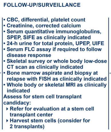
Initial Evaluation
The initial approach to the patient is to evaluate the following parameters:
1. Detection and quantification of a monoclonal (or myeloma) protein (M protein) in the serum or urine, and possible immunoparesis (suppression of the other uninvolved immunoglobulins).
2. Detection of more than 10% of plasma cells on a bone marrow examination, along with flow cytometry, cytogenetics, and fluorescence in situ hybridization testing.
3. Detection of lytic bone lesions or generalized osteoporosis in skeletal x-rays, or whole-body or spinal and pelvic magnetic resonance imaging scans, or focal bone lesions on positron emission tomography-computed tomography scan.
4. Presence of soft tissue plasmacytomas.
5. Serum albumin and beta-2-microglobulin levels.
6. Detection of free kappa and free lambda serum immunoglobulin light chain, with calculation of the serum-free light-chain (FLC) ratio.
7. Presence of hypercalcemia.
8. Detection of renal dysfunction attributable to the plasma cell dyscrasia (induced by gammopathy or amyloidosis.)
9 .Presence of anemia.
10. Presence of circulating plasma cells.
Treatment selection is influenced by the age and general health of the patient, previous therapy, and the presence of complications of the disease.
Classical Definition (CRAB)
• HyperCalcemia
• Renal Insufficiency
• Anemia
• Bone Disease
Expanded definition
• BMPC >= 60%
• >1 PET/MRI lesions
• FLC ratio > 100
Predicts an 80% or more risk of progression in 2 years
Revised International Staging System (R-ISS)
| Stage | Criteria | Median Survival (mo) |
| I | Beta-2-microglobulin <3.5 mg/L and albumin ≥3.5 g/dL | Not reached |
| II | Not R-ISS I or III | 83 |
| III | Beta-2-microglobulin ≥5.5 mg/L and either high LDH or high-risk chromosomal abnormalities by I-FISH (defined as presence of del (17p) and/or translocation t(4;14) and/or translocation t(14;16)) | 43 |
I-FISH = interphase fluorescence in situ hybridization; R-ISS = Revised International Staging System.
Risk Groups for Multiple Myeloma
| Risk Group | Cytogenetic Findings | Disease Characteristics | Median Survival (y) |
| Good risk | Has any of the following cytogenetic findings: (1) no adverse FISH or cytogenetics, (2) hyperdiploidy, (3) t(11;14) by FISH, or (4) t(6;14) by FISH. | These patients most often have (1) disease that expresses IgG kappa monoclonal gammopathies and (2) lytic bone lesions. | 8–10 |
| Intermediate risk | t(4;14) by FISH | These patients often have IgA lambda monoclonal gammopathies and less bone disease. | 5 |
| High risk | Has any of the following cytogenetic findings: (1) del 17p by FISH, (2) t(14;16) by FISH, (3) t(4;14), (4) t(14;20), (5) cytogenetic del 13, (6) nonhyperdiploidy without adverse cytogenetic findings, (7) 1q gain, or (8) plasma cell leukemia. | These patients have (1) disease that expresses IgA lambda monoclonal gammopathies (often) and (2) skeletal-related complications (less often). | <2 |
Treatment Option Overview for Plasma Cell Neoplasms
The major challenge in treating plasma cell neoplasms is to separate the stable, asymptomatic group of patients who do not require immediate treatment from patients with progressive, symptomatic myeloma who may need to be treated immediately.
Monoclonal gammopathy of undetermined significance or smoldering myeloma must be distinguished from progressive myeloma.
Asymptomatic Plasma Cell Neoplasms (Smoldering Multiple Myeloma)
Asymptomatic patients with multiple myeloma who have no lytic bone lesions and normal renal function may be initially observed safely outside the context of a clinical trial.
Increasing anemia is the most reliable indicator of progression.
The following criteria represent the new definition for smoldering myeloma:
• Serum monoclonal protein immunoglobulin (Ig) G or IgA of at least 30 g/L or urinary monoclonal protein of at least 500 mg per 24 hours.
• Clonal bone marrow plasma cells 10% to 60% (>60% represents overt myeloma).
• Absence of amyloidosis or myeloma-defining events as follows:
• Hypercalcemia greater than 1 mg/dl higher than normal.
• Creatinine greater than 2 mg/dl or creatinine clearance less than 40 ml/min.
• Anemia with hemoglobin less than 10.0 g/dl.
• Bone lesions (one or more) on skeletal radiography, computed tomography (CT) or positron emission tomography (PET)-CT.
• Clonal plasma cell percentage in marrow at 60% or more.
• Involved: uninvolved serum-free light chain (FLC) ratio at 100 or more.
• More than one focal lesion of at least 5 mm on magnetic resonance imaging (MRI) of the spine.
A prospective, randomized clinical trial investigated the role of immediate therapy for patients with smoldering multiple myeloma by specifying high-risk patients with both 10% or more marrow plasma cells and a serum monoclonal (or myeloma) protein (M protein) of at least 3 g/dL. The trial randomly assigned 125 patients to receive lenalidomide plus dexamethasone or observation.
• With a median follow-up of 75 months, lenalidomide plus dexamethasone provided benefit in time to progression compared with observation, with a median time not reached (95% confidence interval [CI], 47 months to not reached) compared with 23 months (95% CI, 16‒31 months) (hazard ratio, 0.24; 95% CI, 0.14‒0.41).[Level of evidence: 1iiDiii]
• There was no difference in overall survival (OS) at a median follow-up of 75 months.
• At the beginning of this trial, some of the patients had what would now be considered overt myeloma, based on the updated criteria listed above. This may influence the interpretation of the study because overt myeloma patients might be responsible for some of the benefits seen with therapy.
lenalidomide plus dexamethasone regimen:
lenalidomide 25 mg/day p.o. days 1-21
dexamethasone 40 mg/day p.o. days 1,8,15,21
Every 28 days for 6 cycles
Symptomatic Plasma Cell Neoplasms
Patients with symptomatic advanced disease require treatment.
Treatment most often is directed at reducing the tumor cell burden and reversing any complications of disease, such as renal failure, infection, hyperviscosity, or hypercalcemia, with appropriate medical management. The International Myeloma Working Group (IMWG) has published new criteria for identifying patients with active myeloma who require therapy. These criteria include the following:
• Amyloidosis.
• Hypercalcemia greater than 1 mg/dl higher than normal.
• Creatinine greater than 2 mg/dl or creatinine clearance less than 40 ml/min. Myeloma can cause renal dysfunction via hypercalcemia, amyloidosis, or light chain deposition disease.
• Anemia with hemoglobin less than 10.0 g/dl.
• Bone lesions (one or more) on skeletal radiography, whole-body MRI or spine and pelvis MRI, or PET-CT scans.
• Clonal plasma cell percentage in marrow at 60% or more.
• Involved: uninvolved serum-FLC ratio at 100 or more.
• More than one focal lesion of at least 5 mm on skeletal bone survey, or if negative, total-body MRI, or MRI of the spine and pelvis, or PET-CT scan.
Response criteria have been developed for patients on clinical trials by the IMWG. A very good partial response (VGPR) is defined as a reduction of 90% or more in the serum monoclonal protein and a 24-hour urine monoclonal protein of less than 100 mg. Although not incorporated in the IMWG criteria, many trials report near complete response (nCR) when patients have less than 5% bone marrow plasma cells and unmeasurable serum monoclonal proteins but still have positive serum and/or urine immunofixation. Note that these nCR patients are incorporated into the VGPR group by the IMWG. Patients who achieve a CR by IMWG criteria (with a negative immunofixation along with the clear marrow and unmeasurable serum monoclonal proteins) are often said to have attained a stringent CR if they also normalize their free kappa/lambda light–chain levels and ratio. The clinical utility of these various categories must be validated in clinical trials. Whether these response definitions will translate into clinically meaningful endpoints, such as OS, remains to be seen.
Current therapy for patients with symptomatic myeloma can be divided into the following categories:
• Induction therapies.
• Consolidation therapies, which are less applicable for the very elderly.
• Maintenance therapies.
• Supportive care, such as bisphosphonates.
NCCN Clinical Practice Guidelines: Multiple Myeloma
Initial Diagnostic Workup
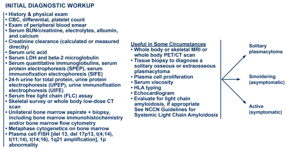
Smoldering Multiple Myeloma
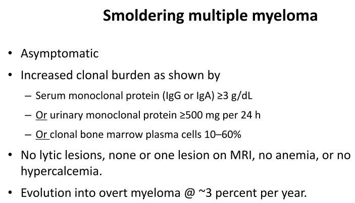
Symptomatic or Active Myeloma
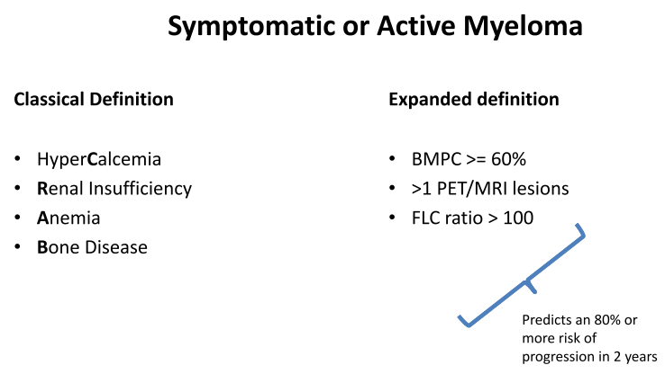
Definition of Multiple Myeloma (Smoldering and Active)
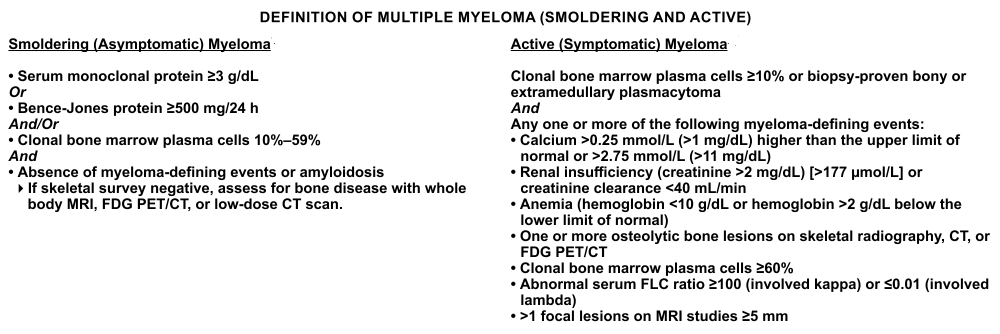
Treatmant for Smoldering (Asymptomatic) Myeloma
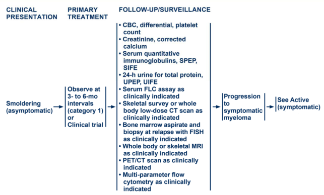
Treatmant for Active (Symptomatic) Myeloma
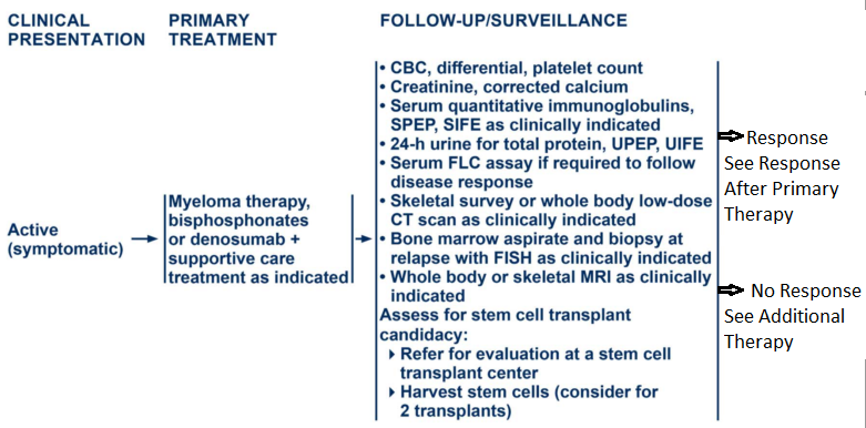
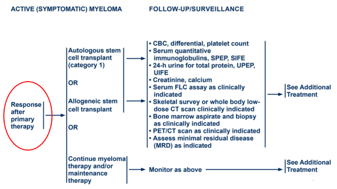
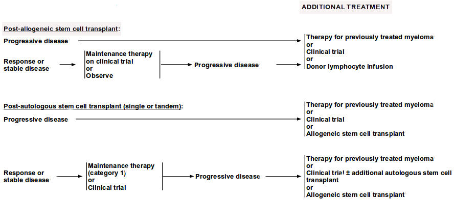
Treatmant for Relapse or Progressive disease Myeloma
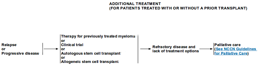
Myeloma Therapy
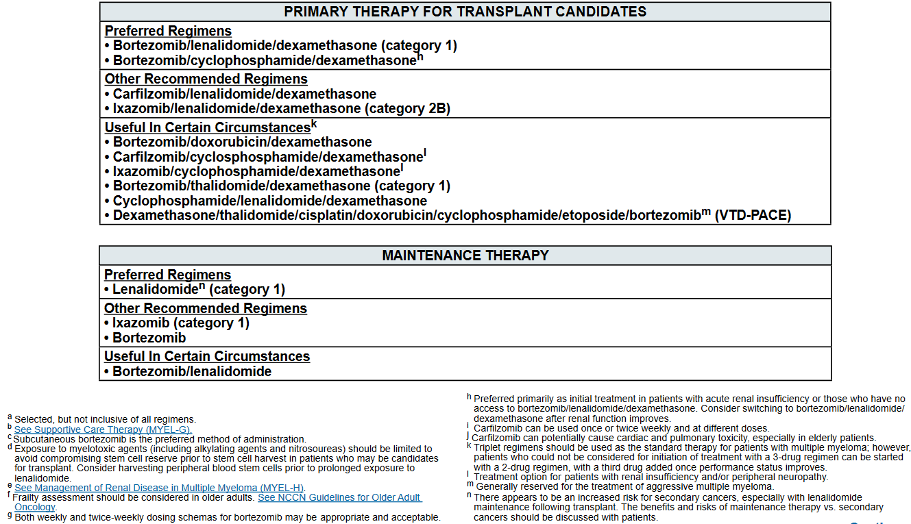
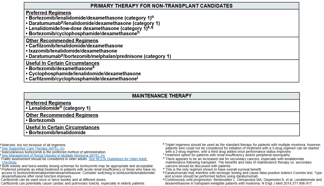
Therapeutic Overview
In fit patients, triple-drug regimens that include bortezomib are considered standard treatment in the absence of a clinical trial. The most commonly used triplets include:
• VRd: lenalidomide + bortezomib + dexamethasone.
• CyBorD: cyclophosphamide + bortezomib + dexamethasone.
Guidelines for choosing induction therapy
Until results become available, outside the context of a clinical trial, clinicians may choose induction therapy based on the following guidelines:
1. In patients younger than 70 years, alkylators are avoided up front to avoid stem cell toxicity with subsequent risks for cytopenias, secondary malignancies, or poor stem cell harvesting if transplantation is considered for consolidation therapy.
2. Bortezomib and/or lenalidomide is combined with dexamethasone for at least 8 months or until best response if consolidation therapy is planned. (Refer to the Lenalidomide and Bortezomib sections of this summary for more information; also refer to the Therapeutic Overview section of this summary for a discussion about the controversy of using doublets or triplets.)
3. The choice of bortezomib or lenalidomide is based on side-effect profile and route of administration.
• Bortezomib is given subcutaneously and can cause neuropathic toxicities. Bortezomib is preferred in the setting of renal impairment.
• Lenalidomide is given orally and can cause an increased risk for deep venous thrombosis (DVT), requiring additional prophylactic medication.
4. Patients with standard-risk disease, as defined in the Stage Information About Plasma Cell Neoplasms section of this summary, might receive induction therapy alone, followed by careful observation after best response. (Refer to the Therapeutic Overview section of this summary for a discussion about the controversy of using a risk-adapted approach in some cases.)
5. Patients with high-risk disease might receive triple-drug induction therapy until best response, followed by consolidation therapy with allogeneic or ASCT.
These guidelines require validation by ongoing clinical trials; participation in clinical trials is the preferred choice, when possible.
Therapy for previously treated multiple myeloma
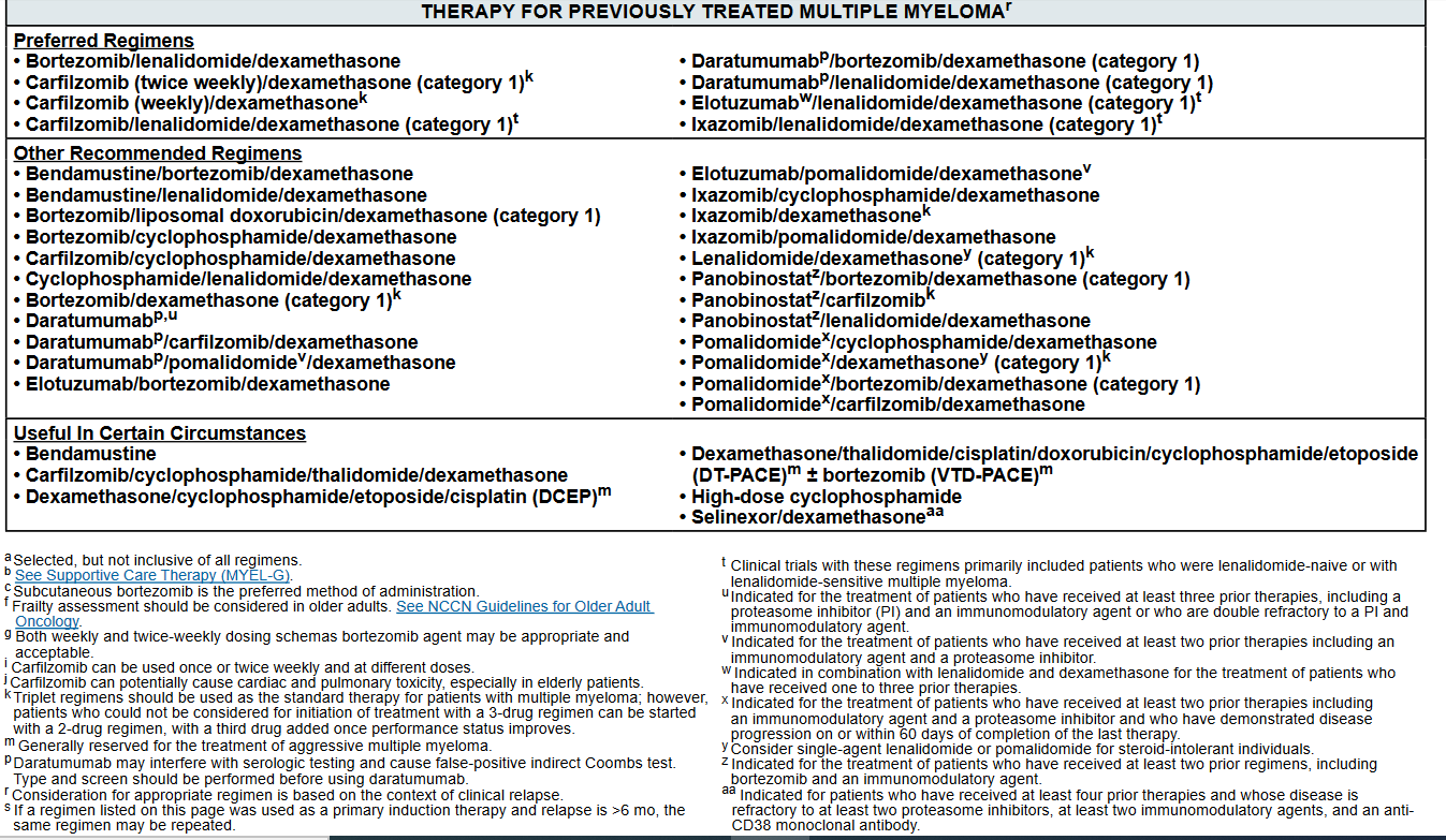
General principles
• Biochemical relapse does not always warrant start of a new therapy
• Duration of initial response defines biology
• Triplet (two active classes + dex) preferred over doublet–At least one drug from a non‐refractory class
• Consider PS, age, comorbidities when selecting therapy
• Take into account prior toxicities/ residual toxicities
• Treat to maximum response and maintain on one drug until progression or tolerability
Not refractory to lenalidomide OR bortezomib
Many options:
• Consider bortezomib, lenalidomide, dex (VRd)
• Repeat induction regimen
• Any of the triplets studied above
VCd is another choice
Refractory to lenalidomide and bortezomib
• Daratumu
• Carfilzomib, pomalidomide, Dex
• Daratumu, pomalidomide, Dex
• Salvage HDT
• Chemotherapy drugs
Alkylators (Melphalan, cyclophosphamide, bendamustine)
Anthracycline
Cisplatin
Etoposide
BCNU
Conclusions
• Ideal initial treatment is the combination of a proteasome inhibitor and an IMiD, or include at least one
• In older patients, frail patients, Len‐Dex is a reasonable choice
• Dose modifications should be done based on patient age and frailty
• SCT has an important role and should be considered early in all eligible patients
• Post‐ASCT maintenance is standard of care
• Bone anti‐resorptive therapy are integral part of supportive care, as is management of infections and toxicity
2018 Updates to Treatment Options for Newly Diagnosed MM
For treatment of newly diagnosed transplant-eligible patients, bortezomib-based triple-drug regimens listed as preferred options include bortezomib/lenalidomide/dexamethasone (VRd) and bortezomib/cyclophosphamide/dexamethasone (VCd).
After VRd demonstrated tolerability and efficacy in patients with newly diagnosed MM in phase II studies, the pivotal phase III multicenter SWOG S07775 trial compared it with lenalidomide/dexamethasone.
In this trial, newly diagnosed patients with MM were randomly assigned to receive 6 months of primary therapy with either VRd or lenalidomide/dexamethasone, each followed by maintenance therapy with lenalidomide/dexamethasone until progression or unacceptable toxicity.
The group treated with VRd showed a significantly longer progression-free survival (PFS) of 43 versus 30 months (hazard ratio [HR], 0.712; 95% CI, 0.56–0.906) and improved median overall survival (OS) of 75 versus 64 months (HR, 0.709; 95% CI, 0.524–0.959).
Based on the significant improvement in PFS and OS seen with VRd, the NCCN panel included this regimen as a category 1, preferred option for the primary treatment of transplant-eligible and non–transplant-eligible patients with newly diagnosed MM.
For patients receiving lenalidomide-based regimens, the panel recommends harvesting peripheral blood stem cells before prolonged exposure to lenalidomide.
Full-dose aspirin is recommended as thromboprophylaxis, and therapeutic anticoagulation is recommended for those at high risk for thrombosis.
The NCCN panel recommends herpes prophylaxis in patients receiving bortezomib therapy (or other PIs) and those receiving anti-CD38 or elotuzumab monoclonal antibody therapy.
Subcutaneous administration is the preferred route for bortezomib, based on the findings of the MMY-3021 trial showing that subcutaneous single-agent bortezomib had noninferior efficacy to intravenous bortezomib with regard to overall response rate (ORR) after 4 cycles. Although time to progression and OS were similar in both groups, patients receiving bortezomib subcutaneously experienced a significant reduction in peripheral neuropathy (PN) (MYEL-D, 1 and 2 of 3; pages 13 and 14).
Data from phase II studies have demonstrated the tolerability and efficacy of VCd in the management of patients with newly diagnosed MM. The NCCN MM Panel has included combination VCd in the list of primary treatments for both transplant-eligible and non–transplant-eligible patients with newly diagnosed MM; this is a preferred option, especially in patients with acute renal insufficiency. According to the panel, patients can consider switching to VRd after renal function improves (MYEL-D, 1 and 2 of 3; pages 13 and 14).
In addition to these bortezomib-based regimens, lenalidomide/low-dose dexamethasone is included as a category 1, preferred regimen for non–transplant-eligible patients.
The international multicenter FIRST trial compared the efficacy and safety of lenalidomide/dexamethasone given continuously or for a fixed duration (72 weeks) with melphalan/prednisone/thalidomide (MPT) in elderly non–transplant-eligible patients with newly diagnosed MM (n=1,623). After a median follow-up of 37 months, risk of progression or death was reduced by 28% in patients receiving continuous lenalidomide/dexamethasone versus MPT (HR, 0.72; 95% CI, 0.61–0.85; P<.001).
An OS benefit was also seen in the lenalidomide/dexamethasone arm versus MPT (HR, 0.78; CI, 0.64–0.96; P=.02). Continuous lenalidomide/dexamethasone also reduced the risk of progression or death compared with 18 cycles of lenalidomide/dexamethasone (HR, 0.70; 95% CI, 0.89–1.20; P=.70), and demonstrated longer median OS.
Results of the ECOG E4A03 trial, which included elderly patients with MM, also demonstrated that lenalidomide/low-dose dexamethasone is a well-tolerated and effective regimen for transplant-eligible and non–transplant-eligible patients.
In this study, the OS rate was significantly higher with lenalidomide/low-dose dexamethasone compared with lenalidomide/high-dose dexamethasone. The inferior survival outcome observed with high-dose dexamethasone was greatest in patients aged ≥65 years.
Patients who did not proceed to autologous stem cell transplant (autoSCT) had an OS rate of 91% with lenalidomide/low-dose dexamethasone at the end of 2 years. The 3-year OS of patients who received 4 cycles of primary treatment with either dose followed by autoSCT was 92%, suggesting that lenalidomide and dexamethasone is a reasonable choice for primary therapy before SCT. However, it should be noted that the choice to proceed to SCT was not randomized, but rather based on physician and patient preference.
Based on the this evidence, lenalidomide/low-dose dexamethasone is listed as a category 1, preferred option in the NCCN Guidelines for non–transplant-eligible patients, especially those who are frail or elderly with standard-risk features.
For transplant-eligible patients, lenalidomide/dexamethasone is listed as a category 1 option in the category “useful in certain circumstances,” with a note that triple-drug regimens are preferred as primary therapy for transplant-eligible patients, although elderly or frail patients may be treated with doublet regimens (MYEL-D, 2 of 3; page 14).
The full list of recommended regimens for transplant-eligible and non–transplant-eligible candidates with newly diagnosed MM can be found on pages 13 and 14 (MYEL-D, 1 and 2 of 3).
Updates to Maintenance Therapy Recommendations
In transplant-eligible patients, multiple phase III randomized trials have evaluated maintenance therapy with lenalidomide after autoSCT. A recent meta-analysis of 1,208 patients randomized to lenalidomide maintenance or placebo showed improved median PFS associated with lenalidomide maintenance (52.8 vs 23.5 months; HR, 0.48; 95% CI, 0.41–0.55).18 At 7 years, OS was 62% in the lenalidomide maintenance group versus 50% for placebo or observation.
In those with high-risk cytogenetics and International Staging System (ISS) stage III disease, a PFS benefit was seen with lenalidomide maintenance versus placebo, but not an OS benefit.
In non–transplant-eligible patients, several randomized trials have explored the benefit of maintenance therapy with lenalidomide after completion of primary therapy.
Data from the phase III MM015 study show that lenalidomide maintenance after melphalan/prednisone/lenalidomide primary therapy significantly reduced the risk of disease progression and increased PFS.
In the FIRST trial, use of lenalidomide until disease progression was associated with superior PFS compared with a fixed duration of lenalidomide.
Updated survival analysis from the CALGB 100104 (Alliance) study at a median follow-up of 91 months reported a median time to progression of 57.3 months (95% CI, 44.2–73.3) with lenalidomide and 28.9 months (95% CI, 23.0–36.3) with placebo (HR, 0.57; 95% CI, 0.46–0.71; P<.0001).
Several reports show higher incidences of secondary malignancies when lenalidomide is used as maintenance therapy post-SCT or in a melphalan-containing regimen. However, there seems to be an increased risk for secondary cancers, especially post-SCT or after treatment with melphalan-containing regimens. According to the results of the FIRST trial, in the continuous lenalidomide/dexamethasone arm, absence of the alkylator melphalan is associated with a lower incidence of second malignancies. In the recent meta-analysis, at a median follow-up of 79.5 months before disease progression on lenalidomide maintenance, the rates of second primary hematologic and solid tumor malignancies were 5.3% and 5.8%, respectively. The benefits of improved PFS with lenalidomide maintenance must be weighed against the increased rate of severe (grade 3 and 4) neutropenia, risk of second cancers, and other toxicities, including cost. The NCCN panel notes that the benefits and risks of maintenance therapy with lenalidomide, including the risk secondary cancers, should be discussed with patients (MYEL-D, 2 of 3; page 14).
Given the significant improvement in PFS and the OS benefit, the NCCN panel now lists single-agent lenalidomide as a category 1, preferred maintenance regimen (MYEL-D, 2 of 3; page 14).
Maintenance with bortezomib has also been evaluated in randomized trials.
Results from the HOVON study show that maintenance with single-agent bortezomib after autoSCT is well tolerated and associated with improvement in ORR.
A multicenter phase III trial showed that consolidation with bortezomib after autoSCT improved PFS only in patients not achieving at least a very good partial response (VGPR) after autoSCT; there was no difference in PFS in patients with a VGPR or better (≥VGPR) after autoSCT. Preliminary results of the phase III UPFRONT study also show that maintenance with single-agent bortezomib is well tolerated when administered after treatment with bortezomibbased primary therapy. Results show that the response rates, including complete response and ≥VGPR, improved after bortezomib maintenance in all arms, with no concomitant increase in the incidence of PN. Bortezomib is listed as an “Other Recommended” maintenance regimen in the NCCN Guidelines (MYEL-D, 2 of 3; page 14).
Updates to Treatment Options for Previously Treated MM
A variety of therapies are available as options for patients with previously treated MM. Choice of therapy depends on the context of clinical relapse, such as prior treatment and duration of response, with options including systemic therapy, autoSCT for eligible patients who did not receive autoSCT as part of their initial treatment, or consideration for a clinical trial.
For those who received autoSCT as part of initial treatment and achieved a durable response or stable disease, consideration should be given to a second SCT on or off clinical trial at the time of relapse/disease progression. If relapse occurs >6 months after completion of the initial primary therapy, patients may be retreated with the same primary regimen. Several new regimens were included as options for the treatment of R/R MM in the 2018 version of the NCCN Guidelines (MYEL-D, 3 of 3; page 15).
Triple-drug regimens remain standard therapy options for patients with MM; however, the NCCN panel notes that selected patients, such as those who are elderly or frail, may be treated with doublet regimens, and a third drug could be added if/when the patient's condition improves.
Results of the phase III ENDEAVOR trial in patients with R/R MM treated with multiple prior lines of therapy showed improved ORR with carfilzomib (twice weekly)/dexamethasone compared with bortezomib/dexamethasone (77% vs 63%). Further, a 2-fold improvement was observed in median PFS with carfilzomib/dexamethasone versus bortezomib/dexamethasone (18.7 vs 9.4 months; HR, 0.53; P<.0001). Additionally, a lower incidence of PN but an increased frequency of heart failure, acute renal failure, and hypertension was seen in the carfilzomib group. OS analysis showed that the carfilzomib/dexamethasone group lived 7.6 months longer (median OS, 47.6 months for carfilzomib vs 40 for bortezomib; HR, 0.791; 95% CI, 0.648–0.964; P=.010).
A phase II study in patients with R/R MM (n=104) evaluated safety and efficacy of weekly dosing of carfilzomib (70 mg/m2) with dexamethasone. The observed ORR was 77% (95% CI, 68–85). At 13.6 months, median PFS was 16.2 months (95% CI, 10.2–21.0). Based on the data from the ENDEAVOR trial, the NCCN MM Panel included the combination of carfilzomib (twice weekly) and dexamethasone as a category 1, preferred option for patients with R/R MM.
Carfilzomib (weekly) with dexamethasone is included under “Other Recommended Regimens” for patients with previously treated MM (MYEL-D, 3 of 3; page 15).
A phase II trial comparing the safety and toxicity of carfilzomib/cyclophosphamide/dexamethasone versus VCd in patients who received one prior regimen for R/R MM showed that carfilzomib/cyclophosphamide/dexamethasone is well tolerated, with carfilzomib having a similar toxicity profile to that seen in other trials.
Considering these data, the panel included carfilzomib/cyclophosphamide/dexamethasone under “Other Recommended Option” for patients with previously treated MM (MYEL-D, 3 of 3; page 15).
For patients with R/R MM who have received at least one prior therapy, 3 regimens were added as category 1: ixazomib/lenalidomide/dexamethasone for those who have received at least one prior therapy, and daratumumab/lenalidomide/dexamethasone and daratumumab/bortezomib/dexamethasone based on the results of the multicenter, randomized, phase III POLLUX and CASTOR trials, respectively.
For patients with R/R MM who have received ≥2 prior therapies, including an IMiD and a PI, and who have demonstrated disease progression on or within 60 days of completion of the last therapy, 2 new regimens were added as options in the updated guidelines: ixazomib/pomalidomide/dexamethasone and daratumumab/pomalidomide/dexamethasone. The inclusion of ixazomib/pomalidomide/dexamethasone was based on the results of recently reported phase I/II studies. The inclusion of daratumumab/pomalidomide/dexamethasone was based on the results of a phase Ib multicenter study that included >100 patients who had received ≥2 prior lines of therapy (excluding daratumumab or pomalidomide). At a median follow-up of 13.1 months, the ORR was 60%, and median PFS and OS were 8.8 and 17.5 months, respectively; the estimated survival at 1 year was 66%. Phase III studies of daratumumab/pomalidomide/dexamethasone compared with pomalidomide/dexamethasone are ongoing. The addition of IMiDs to daratumumab in patients with refractory MM may overcome refractoriness in both agents.
Daratumumab can interfere with cross-matching and red blood cell antibody screening; therefore, the NCCN panel recommends performing a type and screen before administering this agent to inform future matching. Daratumumab can also interfere with immunofixation assays used to determine response to therapy if the dominant myeloma clone is IGG kappa, because daratumumab is itself an IGG monoclonal antibody.
Patients with an aggressive relapse may need multidrug combinations for effective disease control; VTD-PACE (bortezomib, dexamethasone, thalidomide, cisplatin, doxorubicin, cyclophosphamide, and etoposide) is one such regimen that is now an option listed under “Useful in Certain Circumstances” for treating previously treated aggressive MM (MYEL-D, 3 of 3; page 15).
The full list of recommended regimens for previously treated MM can be found on page 15 (MYEL-D, 3 of 3).
Conclusions
These NCCN Guidelines Insights highlight important updates/changes specific to treatment options for newly diagnosed and R/R MM in the 2018 version of the NCCN Guidelines for MM.
The NCCN Guidelines are in continuous evolution, and are updated annually or more often if new, high-quality clinical data become available. The recommendations in the NCCN Guidelines, with few exceptions, are based on evidence from clinical trials.
Expert medical clinical judgment is required when applying these guidelines in the context of individual clinical circumstances to provide optimal care.
Both physicians and patients have a responsibility to jointly explore and select the most appropriate option from among the available alternatives. When possible, consistent with NCCN philosophy, the panel strongly encourages patient/physician participation in prospective clinical trials.